In a groundbreaking study conducted by researchers at the Picower Institute for Learning and Memory at MIT, scientists have unveiled unprecedented insights into how the brain’s visual system undergoes rapid and extensive rewiring during a critical developmental window. This research stands out as the first to track the synaptic connections of individual neurons in the visual cortex of live mice continuously over the entire “critical period” when binocular vision—the brain’s ability to integrate input from both eyes—is refined. The findings challenge previous notions about neural stability and demonstrate a far more dynamic landscape of synaptic turnover than ever imagined.
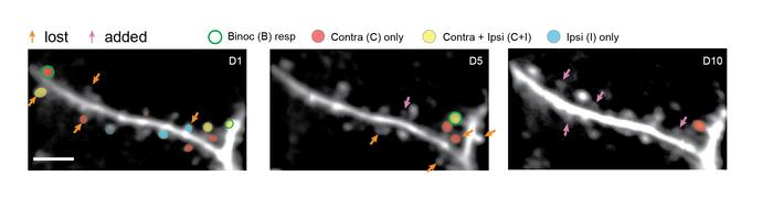
The scientific team focused their attention on dendritic spines—the tiny protrusions on neurons where synapses, or communication points between neurons, reside. By using state-of-the-art imaging techniques, they repeatedly captured images of the same dendrites across ten days during the critical period of visual development. This continuous monitoring revealed that only about 40 percent of the initial spines present on day one persisted by day ten, indicating that the vast majority of synaptic connections are transient and subject to intense remodeling during this phase.
To provoke specific neural responses, the researchers exposed young mice to controlled visual stimuli consisting of black and white grating patterns moving in various orientations and directions. During this exposure, they concurrently recorded both structural changes—appearance and disappearance of dendritic spines—and functional activity of the neurons and their individual spines. Such dual examination allowed the team to correlate physical synaptic turnover with shifts in neuronal response properties, illuminating the mechanisms underlying the maturation of binocular vision.
.adsslot_7BmzDQ8cNH{ width:728px !important; height:90px !important; }
@media (max-width:1199px) { .adsslot_7BmzDQ8cNH{ width:468px !important; height:60px !important; } }
@media (max-width:767px) { .adsslot_7BmzDQ8cNH{ width:320px !important; height:50px !important; } }
ADVERTISEMENT
The extent of synaptic dynamics was staggering. From day one to day five, nearly one-third of the original spines were pruned away, while new spines emerged to replace some of those lost. Between days five and ten, the process continued with similar turnover rates. This continuous cycle of spine elimination and formation ultimately led to the drastic filtering of which synapses remained, sculpting a refined and efficient binocular circuit. Remarkably, only a minority of neurons that initially responded to visual stimuli maintained their responsiveness by day ten, suggesting that neurons may change their functional roles as development proceeds.
A core objective of the study was to decipher what governed the survival or elimination of individual spines during this critical period. The experimental evidence pointed towards two interdependent factors. Firstly, spines that exhibited higher activity levels—meaning they were more frequently involved in neural signaling—were more likely to be sustained. Secondly, the selective retention of spines was biased towards those that aligned with the orientation preference of the parent neuron’s soma. This orientation tuning is crucial for the brain’s ability to detect and interpret edges and shapes in the visual environment, a fundamental aspect of visual perception.
An intriguing observation was that spines responding to inputs from both eyes—termed binocular spines—exhibited greater activity than those responsive to only one eye. This higher activity likely confers a survival advantage within the competitive synaptic landscape, reinforcing the “use it or lose it” principle long hypothesized in neuroscience. Such findings suggest that the brain’s wiring is intricately dependent on functional relevance and synaptic efficacy, rather than mere structural presence.
Another sophisticated phenomenon uncovered was the emergence of functional clusters of activity along dendrites. Neighboring spines began to synchronize their activation patterns during the critical period, effectively amplifying their collective impact on neuronal outputs. This spatial clustering aligns with the concept of heterosynaptic plasticity, where the activity of one synapse affects its neighbors, leading to a cooperative strengthening of functionally related groups of synapses, rather than isolated synaptic modifications.
To further validate these experimentally observed patterns, the researchers constructed computational models simulating neural networks embodying the principles of Hebbian plasticity—where synapses strengthen if they are consistently co-active—and heterosynaptic plasticity. The models reliably reproduced the turnover and refinement of synaptic populations akin to the in vivo observations, reinforcing the notion that these two plasticity mechanisms are essential in shaping the brain’s emerging binocular circuitry.
The implications of these findings ripple through the fields of developmental neuroscience and sensory processing research. They provide concrete empirical support for the intricate balance between synaptic formation, elimination, and functional tuning that underlies perceptual development. Notably, the results elucidate how early sensory experiences can powerfully influence neural circuitry by selectively stabilizing synapses that contribute to meaningful interpretations of the environment while discarding those that are redundant or misaligned.
Beyond foundational science, this research may inform therapeutic strategies for visual disorders implicating synaptic dysfunction or developmental anomalies, such as amblyopia (“lazy eye”). By understanding the precise dynamics and rules guiding synaptic refinement, future interventions could be better tailored to promote beneficial rewiring during or possibly beyond the critical period.
The study was pioneered by former MIT graduate student Katya Tsimring under the leadership of senior author Mriganka Sur, the Paul and Lilah Newton Professor at the Picower Institute. Their team painstakingly tracked 793 dendritic spines across 14 neurons, meticulously quantifying structural changes alongside functional responses. Their integrative approach sets a new standard for in vivo studies of neural development, showcasing how live imaging combined with computational modeling can unravel complex brain processes at an unprecedented resolution.
This compelling body of work was published in the prestigious journal Nature Communications, highlighting the synergy between experimental innovation and theoretical validation. The research was supported principally by the National Institutes of Health, the Picower Institute, and the Freedom Together Foundation, reflecting the critical role of funding agencies in advancing cutting-edge neuroscience.
Looking forward, this study opens avenues for exploring how similar synaptic dynamics might operate in other sensory systems or brain regions, broadening our comprehension of brain plasticity. As the field progresses, the fusion of longitudinal live imaging and sophisticated computational tools promises to elucidate the neural code embedded within synaptic architectures shaping perception, cognition, and behavior.
Subject of Research: Animals
Article Title: Large-scale synaptic dynamics drive the reconstruction of binocular circuits in mouse visual cortex
Web References: 10.1038/s41467-025-60825-y
Image Credits: Sur Lab/MIT Picower Institute
Keywords: Neuroscience, Vision, Developmental neuroscience, Brain development, Synapse formation, Sensory perception, Brain
Tags: binocular vision researchcritical period for visiondendritic spine remodelinglive imaging of neuronsMIT Picower Institute studyneural connections developmentneuroscience of visionsynaptic turnover in neuronstransient synaptic connectionsunderstanding visual developmentvisual cortex dynamicsvisual stimuli impact on brain


