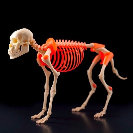
In a groundbreaking advancement for orthopedic medicine, scientists have developed an innovative device that revolutionizes how bone grafts are created and applied during surgical procedures. This state-of-the-art tool, essentially a modified glue gun, can 3D print bone grafts directly onto fractures and defects while a patient is undergoing surgery. Described in the Cell Press journal Device, this pioneering technique holds the promise of expediting the process of bone repair, making surgical interventions more efficient and effective.
Traditionally, bone implants used in surgeries have been made from various materials such as metals, donor bones, or, more recently, 3D-printed materials. The conventional approach necessitates careful pre-surgical planning, where implants must be customized and manufactured before a patient’s surgery. However, in cases involving complex or irregular bone fractures, this preparatory phase can be a significant challenge. In contrast, the new method allows for the direct creation of customizable bone scaffolds tailored to the specific anatomy of the patient, right at the site of injury, eliminating the need for any preoperative fabrication.
Jung Seung Lee, an associate professor of biomedical engineering at Sungkyunkwan University and a co-author of the study, highlights the advantages of this technology. “Our proposed technology offers a distinct approach by developing an in situ printing system that enables real-time fabrication and application. This innovative method allows for highly accurate anatomical matching, particularly beneficial during surgeries involving irregular or complex defects,” he stated. This real-time capability not only simplifies the process for surgeons but also enhances the overall quality of care patients receive during critical procedures.
The filament material powering this device contains two crucial components: hydroxyapatite (HA), a naturally occurring mineral component found in bone, known for promoting healing, and polycaprolactone (PCL), a biocompatible thermoplastic. PCL can be liquefied at temperatures as low as 60°C, allowing it to flow and conform seamlessly to the irregular shapes of fractured bone while remaining cool enough to prevent thermal injury to surrounding tissues during application. By modifying the proportion of HA to PCL in the filament, the research team can customize the strength and hardness of the grafts to match the varied anatomical requirements presented in patients.
The surgeon’s ability to manipulate the device manually grants them unprecedented control during the printing process. This capability ensures that the grafts can be accurately placed in precise orientations, directions, and depths according to the unique characteristics of the patient’s injury. Lee noted that the entire printing process could be completed in a matter of minutes, significantly reducing overall operative times. This efficiency becomes critical in surgical environments, where time limitations often dictate the quality of care in emergency situations.
One of the common pitfalls of surgical implants is the heightened risk of postoperative infections. Acknowledging this concern, the researchers ingeniously included two powerful antibacterial agents, vancomycin and gentamicin, into the filament material used for 3D printing the grafts. Experiments conducted both in petri dishes and liquid mediums have shown promising results, with the filament scaffolds effectively inhibiting the growth of notorious bacteria such as E. coli and Staphylococcus aureus. Notably, the release of these drugs is sustained, allowing them to diffuse directly to the surgical site over several weeks, thereby reducing the patient’s risk of infection without the drawbacks associated with systemic antibiotic use.
This localized delivery system is poised to bring significant clinical advantages. By minimizing the side effects and mitigating the risk of developing antibiotic resistance associated with broader systemic treatments, this innovative approach enables targeted protection against infections. Lee emphasizes the implications this could have for patients undergoing surgeries involving implants, where infection rates are a primary concern.
To demonstrate the efficacy of this technology, the research team conducted proof-of-concept tests on rabbits with severe femoral bone fractures. Remarkably, within 12 weeks of surgery, the results indicated no signs of infection or tissue necrosis. The implants demonstrated substantial bone regeneration compared to traditional bone cement, a common material utilized for addressing similar injuries in clinical settings.
The integrated scaffold is designed to carry out two functions: biological integration with the surrounding bone tissue and gradual degradation over time. Specifically, it is crafted to be substituted by newly formed bone as healing progresses. Lee and his team observed that in comparisons with previous grafts, their printed scaffolds yielded superior outcomes in essential structural metrics such as bone surface area and cortical thickness, correlating to improved healing and integration outcomes.
On the horizon, the research team plans to enhance the antibacterial properties of their 3D-printed scaffolds further and prepare for human clinical trials. Lee encapsulates the future vision succinctly: “For clinical adoption, our approach will first necessitate the development of standardized manufacturing protocols, validated sterilization procedures, and preclinical studies conducted in larger animal models to satisfy regulatory requirements.” If these benchmarks can be met successfully, the team is optimistic that this technology will transform bone repair practices directly within the operating room.
The innovative device represents a significant leap forward in medical technology, promising to alter how bone injuries are treated in real-time during surgical operations. As this research progresses and human trials commence, the potential for widespread clinical application could lead to higher success rates in bone repair, ultimately improving the quality of life for countless patients recovering from traumatic injuries.
This remarkable development serves as a true testament to the evolving landscape of biomedical engineering and the impact that interdisciplinary collaboration can have on improving patient outcomes in modern medicine.
Subject of Research: Animals
Article Title: In situ printing of biodegradable implant for healing critical-sized bone defect
News Publication Date: 5-Sep-2025
Web References: http://www.cell.com/device/home
References: 10.1016/j.device.2025.100873
Image Credits: Jeon et al. / Device
Keywords
Biomedical engineering, Additive manufacturing, Bone fractures, Traumatic injury, Bones, Medical technology, Regenerative medicine
Tags: 3D printing bone graftsadditive manufacturing in healthcareanimal models in orthopedic researchbiomedical engineering breakthroughscustomizable bone scaffoldsdirect application bone graftingefficient surgical interventionsfracture treatment advancementsorthopedic medicine innovationspatient-specific bone implantsrevolutionary medical devicessurgical bone repair technologies


