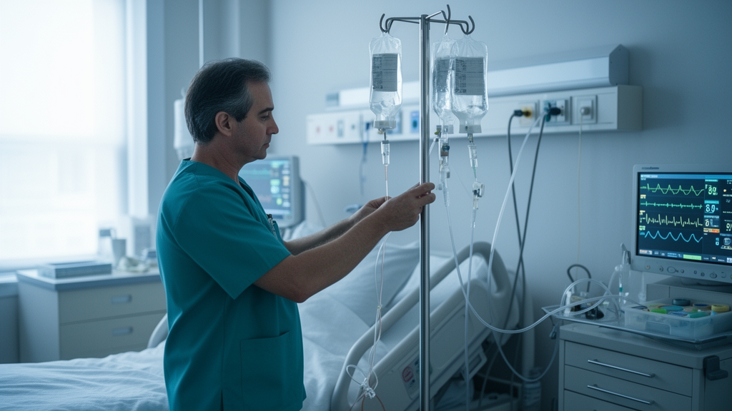
A review published in Annals of Intensive Care examines the physiological pathways by which intravenous crystalloid fluid distributes throughout the human body, aiming to clarify why fluid overload can lead to severe complications in critically ill patients. Authored by Robert G. Hahn, the article synthesizes current knowledge on fluid kinetics to explain the pathophysiological mechanisms behind overhydration, a condition where both excessive and insufficient fluid volumes can promote adverse outcomes. The review highlights that liberal fluid administration is often necessary to maintain tissue perfusion in life-threatening conditions, yet the precise distribution of this fluid remains a complex interplay of vascular and interstitial dynamics.
The primary locations for excess crystalloid fluid in humans are identified as the skin, intestinal wall, and lungs. These tissues are selected for fluid accumulation because they possess high compliance, meaning they can expand easily to accommodate additional volume. In contrast, bone tissue does not undergo volume expansion, and organs surrounded by tight capsules accumulate minimal excess fluid. The interstitial volume in the skin accounts for 50% of its mass, whereas in muscle it accounts for only 10%, and the compliance for volume expansion in skin is twice that of muscle.
Volume kinetic analysis reveals that the human body contains three distinct extracellular fluid compartments that expand in response to electrolyte-based crystalloid infusion. The first is the plasma volume, which initially receives the infused fluid. The second is a fast-exchange interstitial fluid space, referred to as Vt1, which equilibrates with the plasma within approximately 30 minutes. The third is a slow-exchange interstitial space, known as Vt2 or the “third fluid space,” which acts as an overflow reservoir. Vt2 opens for volume expansion only after Vt1 has increased by 600 to 800 mL, a threshold typically reached by infusing 1.3 to 1.5 liters of crystalloid fluid over 30 minutes.
The opening of the slow-exchange space is a sudden event that likely corresponds to the point where interstitial hydrostatic pressure rises from its normal sub-atmospheric range to zero. Before this threshold is crossed, water molecules may enter Vt2 but are quickly exchanged due to low compliance. Once the threshold is exceeded, the sub-atmospheric pressure that normally holds tissues together is lost, leading to the formation of scattered fluid pools between cell bundles. This phenomenon is clinically observed as pitting edema in the skin and can also occur in the heart muscle, posing significant risks during severe overhydration.
Morphological studies in animals provide evidence of the tissue damage associated with extreme fluid overload. In rabbits, the infusion of 100 mL/kg of saline over 30 minutes caused interstitial dilatation and swelling of the heart, along with hypoxic lesions in the left ventricle and papillary muscles. In mice, higher doses of saline resulted in hypoxic changes in the heart and lesions of the histoskeleton, with mortality rates reaching 30% for both 200 mL/kg and 300 mL/kg doses. Although these findings demonstrate infusion-related lesions in experimental animals, the occurrence of similar morphological damage in overhydrated humans remains poorly documented, highlighting a gap in current clinical understanding.
General anesthesia significantly alters fluid kinetics by inhibiting the diuretic response to plasma volume expansion by 85 to 90%. This inhibition is largely due to a reduction in arterial pressure, which is transmitted via renal sympathetic fibers. Additionally, anesthetic agents inhibit lymphatic pumping, slowing the return of distributed fluid to the plasma. Despite lower arterial pressure, capillary filtration accelerates under general anesthesia due to the relaxation of pre-capillary sphincters, which increases the surface area available for transcapillary filtration. The combined effect of these changes is greater fluid retention in the whole body, with a disproportionately larger increase in interstitial volume compared to plasma volume.
In inflammatory conditions such as sepsis and preeclampsia, the dynamics of fluid distribution are further complicated by the presence of pro-inflammatory cytokines. Molecules such as TNF-alpha, IL-1-beta, and IL-6 reverse integrin-collagen bindings in the interstitium, decreasing interstitial pressure and creating a suction effect that withdraws fluid from the fast-exchange space into the slow-exchange space. This process reduces lymphatic flow, leading to a triad of hypovolemia, hypoalbuminemia, and peripheral edema. In septic sheep, this competition between fluid accumulation in Vt2 and lymphatic flow resulted in an abnormal ratio of return flow to third-space accumulation, suggesting that lymphatic flow could stagnate if crystalloid fluid is not administered early in the course of sepsis.
The slow-exchange space is not a bottomless reservoir; its capacity is limited by the compliance of the interstitial tissue. Re-analysis of experiments in healthy volunteers suggests that acute filling of Vt2 peaks at approximately 600 mL. Further volume loading beyond this point expands the fast-exchange space rather than the slow-exchange space. However, longstanding overhydration increases the compliance of both vascular and interstitial spaces over time, a process known as stress relaxation. This delayed compliance allows Vt2 to gradually accommodate more fluid during prolonged intensive care stays, explaining why body weight increases after major surgery can persist for up to one week.
Strategies to manipulate the slow-exchange space include infusing less fluid or providing it more slowly to allow the kidneys time to excrete excess volume. Hyper-oncotic albumin is another approach aimed at modifying peripheral edema. Data from healthy volunteers show that 20% albumin withdraws 3.4 times the infused volume from the interstitium within five hours, primarily via the lymphatic route. This recruitment of interstitial fluid stimulates diuresis, with two-thirds of the withdrawn volume excreted as urine. The intravascular persistence time of the expanded volume is longer in postoperative and surgical patients compared to healthy volunteers, which is a critical consideration when planning repeat infusions.
Manual compression has also been evaluated as a method to reduce total body edema in septic patients, with studies showing increased cardiac preload and reduced wrist circumference in intensive care settings. The interstitium is normally under a constant state of dehydration, and counter-pressure prevents the swelling that tissues undergo when exposed to isotonic saline in vitro. Effective lymphatic flow coupled with slow capillary filtration is required to maintain the sub-atmospheric pressure of the interstitium, which is essential for the eventual closure of the slow-exchange space. Restoration of this pressure is a slow process that may require days for full completion, even after the initial fluid overload has been addressed.
The review concludes that while the physiological mechanisms of fluid distribution are becoming better understood through volume kinetic analysis, the pathophysiological mechanisms explaining the complications and fatal outcomes of overhydration in humans remain insufficiently known. The slow-exchange interstitial space operates as a critical overflow reservoir in settings of volume overload, and its interaction with inflammatory processes significantly impacts patient outcomes.
Fluid administration is a standard component of intensive care, where water loss from evaporation, estimated at approximately 700 mL per day, must be compensated if patients cannot eat or drink. Maintaining a urine flow exceeding 0.5 mL per minute is also necessary to ensure the full excretion of metabolic end products. In severe disease, blood flow redistribution occurs due to blunted adrenergic tonus and subsequent vasodilation, requiring plasma volume expansion with electrolyte-based crystalloid or colloid fluids to preserve organ function. This resuscitation typically lasts one to three days, followed by a de-resuscitation phase when the patient’s condition improves. However, positive fluid balance is associated with increased mortality in intensive care units, and liberal administration during surgery can prolong hospital stays or necessitate intensive care admission.
The distribution of excess fluid is governed by tissue compliance, which varies significantly across the body. Consequently, most excess fluid accumulates in the skin, potentially impairing wound healing and exacerbating local infections. The lungs are another primary site for fluid accumulation, where volume overload promotes pulmonary edema and acute respiratory distress syndrome, which can develop with a delay of several days. The gastrointestinal wall also accumulates excess fluid, as its fenestrated capillaries allow for rapid transfer of fluid and solutes. Both animal and human studies indicate that the gastrointestinal wall retains more water when infused with crystalloid rather than colloid fluid.
Clinical observations correlate these anatomical sites with specific complications. Administration of more than 2 liters of crystalloid has been linked to prolonged postoperative gastrointestinal recovery, while infusing 6 liters instead of 3 liters during colon surgery increased the risk of wound infection, suture insufficiency, bleeding, and pneumonia. In a separate observation, rapid administration of 2 liters of Ringer’s solution over 15 minutes in young women caused acute swelling sensations in the face and arms, along with slight dyspnea. These symptoms did not occur when the infusion time was doubled, suggesting that tissues require more time to adapt to volume changes.
Subject of Research: Physiological distribution of crystalloid fluid and mechanisms of overhydration in intensive care
Article Title: Where does the fluid go?
Article References: Hahn, R. G. (2025). Where does the fluid go?. Annals of Intensive Care, 15(1), Article 156. https://doi.org/10.1186/s13613-025-01579-0
Image Credits: AI Generated
DOI: 10.1186/s13613-025-01579-0
Keywords: fluid resuscitation, interstitial edema, volume kinetics, intensive care, crystalloid infusion, lymphatic flow, sepsis, preeclampsia, albumin therapy, pathophysiology, does, fluid
News Source: Ophelia Keating. (November 10, 2026). Review Maps Fluid Distribution and Overload Risks in Intensive Care. Scienmag.