
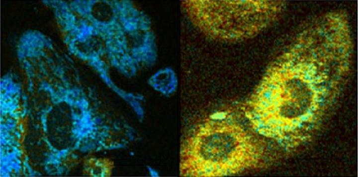
Credit: Irene Georgakoudi, Tufts University
MEDFORD/SOMERVILLE, Mass. (March 7, 2018) — Metabolic changes in cells can occur at the earliest stages of disease. In most cases, knowledge of those signals is limited, since we usually detect disease only after it has done significant damage. Now, a team led by engineers at Tufts University School of Engineering has opened a window into the cell by developing an optical tool that can read metabolism at subcellular resolution, without having to perturb cells with contrast agents, or destroy them to conduct assays. As reported today in Science Advances, the researchers were able to use the method to identify specific metabolic signatures that could arise in diabetes, cancer, cardiovascular and neurodegenerative diseases.
The method is based on the fluorescence of two important coenzymes (biomolecules that work in concert with enzymes) when excited by a laser beam. The coenzymes – nicotinamide adenine dinucleotide (NADH) and flavin adenine dinucleotide (FAD) – are involved in a large number of metabolic pathways in every cell. To find out the specific metabolic pathways affected by disease or stress, the Tufts scientists looked at three parameters: the ratio of FAD to NADH, the fluorescence "fade" of NADH, and the organization of the mitochondria as revealed by the spatial distribution of NADH within a cell (the energy producing "batteries" of the cell).
The first parameter – the relative amounts of FAD to NADH – can reveal how well the cell is consuming oxygen, metabolizing sugars, or producing or breaking down fat molecules. The second parameter – the fluorescence "fade" of NADH – reveals details about the local environment of the NADH. The third parameter – the spatial distribution of NADH in the cells – shows how the mitochondria split and fuse in response to cellular growth and stress.
"Taken together, these three parameters begin to provide more specific, and unique metabolic signatures of cellular health or dysfunction," said Irene Georgakoudi, Ph.D., corresponding author of the study and a professor of biomedical engineering in the School of Engineering at Tufts. "The power of this method is the ability to get the information on live cells, without the use of contrast agents or attached labels that could interfere with results."
Other methods exist for non-invasively tracking the metabolic signatures of disease, such as the PET scan, which is often used in research. But while PET scans provide low resolution information with excellent depth penetration into living tissues, the optical method introduced by the Tufts researchers detects metabolic activity at the resolution of single cells, although mostly near the surface.
That is not necessarily a limitation. Many diseases can be detected at the surface of tissues, including cancer, while many pre-clinical studies are performed with animal models and engineered three-dimensional tissues that can benefit from being monitored non-destructively. The method developed by Georgakoudi and colleagues may prove to be a powerful research tool for understanding their metabolic signatures.
###
Other authors on the paper are: lead author Zhiyi Liu, Dimitra Pouli, Carlo Alonzo, Antoine Varone, all of the Department of Biomedical Engineering at Tufts University; Kyle Quinn, formerly of the Department of Biomedical Engineering and now at the University of Arkansas; Sevasti Karaliota and Katia Karalis, of the Biomedical Research Foundation at the Academy of Athens, Greece; and Karl Münger, of the Sackler School of Graduate Biomedical Sciences at Tufts.
This work was supported by the National Institutes of Health (NIH R21EB019079, NIH K99EB017723 NIH R01CA066980 and R00EB017723) and the American Cancer Society (RSG-09-174-01-CCE). The content is solely the responsibility of the authors and does not necessarily represent the official views of the National Institutes of Health.
Liu, Z., Pouli, D., Alonzo, C.A., Varone, A., Karaliota,S., Quinn, K.P., Münger, K., Karalis, K.P., Georgakoudi, I. "Mapping metabolic changes by noninvasive, multiparametric, high-resolution imaging using endogenous contrast" Sci. Adv. 2018; 4: eaap9302. DOI: 10.1126/sciadv.aap9302
About Tufts University
Tufts University, located on campuses in Boston, Medford/Somerville and Grafton, Massachusetts, and in Talloires, France, is recognized among the premier research universities in the United States. Tufts enjoys a global reputation for academic excellence and for the preparation of students as leaders in a wide range of professions. A growing number of innovative teaching and research initiatives span all Tufts campuses, and collaboration among the faculty and students in the undergraduate, graduate and professional programs across the university's schools is widely encouraged.
Media Contact
Mike Silver
[email protected]
617-627-0545
@TuftsUniversity
http://www.tufts.edu
Related Journal Article
http://dx.doi.org/10.1126/sciadv.aap9302


