Diagnostic imaging is indispensable in healthcare, as it allows clinicians to detect and diagnose a variety of medical conditions. Despite significant advances to imaging technology, however, existing single imaging techniques often fall short of addressing all diagnostic scenarios, which leads to an increased reliance on multiple imaging types and higher healthcare costs. In response to this challenge, researchers at the University of Illinois Urbana Champaign have developed a dual-modality imaging technique that not only delivers comprehensive diagnostic information but also provides a cost-effective solution for healthcare providers.
Credit: The Grainger College of Engineering at the University of Illinois Urbana-Champaign
Diagnostic imaging is indispensable in healthcare, as it allows clinicians to detect and diagnose a variety of medical conditions. Despite significant advances to imaging technology, however, existing single imaging techniques often fall short of addressing all diagnostic scenarios, which leads to an increased reliance on multiple imaging types and higher healthcare costs. In response to this challenge, researchers at the University of Illinois Urbana Champaign have developed a dual-modality imaging technique that not only delivers comprehensive diagnostic information but also provides a cost-effective solution for healthcare providers.
Ultrasound (US) imaging is a prevalent and widely employed diagnostic tool in healthcare. However, it is limited by low image quality and must often be paired with higher quality and more expensive imaging methods, such as MRI. To enhance US imaging, electrical and computer engineering assistant professors Yun-Sheng Chen and Yang Zhao, along with ECE graduate student Shensheng Zhao, integrated photoacoustic (PA) imaging, super-resolution ultrasound imaging, and a sparsity-constrained optimization method to create a dual-modality, super-resolution medical imaging technique. This new research, “Hybrid Photoacoustic and Fast Super-Resolution Ultrasound Imaging”, was recently published in Nature Communications.
Zhao highlights that this new imaging tool features “more accessibility, portability, and cost-effectiveness. This technique is significantly cheaper than clinical medical imaging techniques, while providing similar functions.”
Traditional imaging techniques are most effective at identifying diseases when a patient exhibits structural changes in their organs. Unfortunately, by the time these changes become apparent, the disease has often progressed to an advanced stage. To address this shortcoming, the researchers aimed to develop a more comprehensive imaging technique capable of detecting additional physiological and biochemical abnormalities like changes in blood flow and tissue oxygenation. By incorporating two emerging ultrasound-based imaging approaches, the team hopes to facilitate earlier disease detection, potentially improving patient outcomes and prognosis.
Photoacoustic imaging is a technique that utilizes light from a laser pulse, which is absorbed by tissues in the body, generating ultrasound signals that produce images. Interestingly, each tissue exhibits a unique signature after interacting with the light, allowing both the structure and content of the tissue to be identified. PA imaging can visualize physiological and biochemical processes within the body, such as blood oxygen content, tissue composition, inflammation, and the distribution of imaging agents (functional information). Despite the strengths of PA imaging, it does also have its limitations- its resolution is restricted, rendering it unable to resolve fine structures like microvasculature, which are crucial targets in various diseases, including cancer and kidney disease.
Super-resolution ultrasound imaging employs microbubbles as contrast agents that flow in the bloodstream and produce very strong ultrasound signals. Advanced signal processing is used to eliminate the ultrasound tissue background signal, focusing solely on the signal of the individual imaging agents in the bloodstream. This technique can achieve high resolution, making it ideal for imaging microvasculature and measuring corresponding blood flow. As both super-resolution ultrasound and photoacoustic imaging can share the same ultrasound imaging system, they are well-suited for developing dual-modality imaging.
Combining super-resolution US imaging with PA imaging may sound straightforward, but each technique has its own hardware requirements and acquisition speeds. Differing acquisition speeds are particularly important, as patients’ natural movements, such as their breathing and heartbeat, can cause the imaged information to shift over time. Application of sparsity-constrained optimization to the signal processing aligned the speed of the two imaging techniques, enabling a smooth scan of the imaging area. This approach allowed them to accelerate the frame rate of super-resolution ultrasound imaging by up to 37 times with synthetic data and 28 times with in vivo data. The matched imaging speeds enable interleaved recording of the two imaging types, allowing successful co-registration of chemical composition, blood flow, and structure.
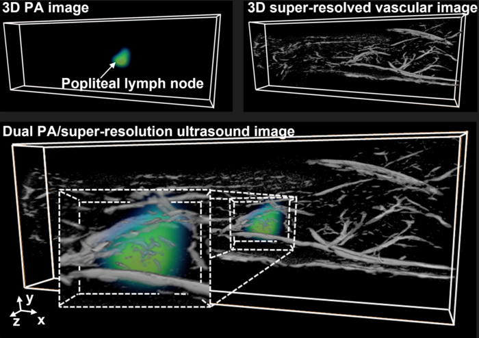
The research team demonstrated their groundbreaking technique in two critical in vivo scenarios—lymph nodes and kidneys. The lymph nodes are among one of the first places cancer will metastasize, and doctors will check a patients lymph nodes to determine if a lymphadenectomy is necessary. For this process, it is important to know where the lymph node is and visualize the complex vasculature surrounding the cancer cells. With current techniques, either the vasculature can be seen, or the lymph node can be located, but not both. This new technique has shown that it can provide information on both the lymph node location and vasculature.
Kidneys are another significant area of interest, as approximately 15% of Americans will develop kidney disease and 40% of those with chronic kidney disease are unaware of their condition until diagnostic imaging is performed. The vasculature and oxygenation of the kidney play a vital role in the development of chronic kidney disease. However, like in the case of lymph nodes, current imaging modalities cannot adequately capture both structural and functional information. The dual imaging modality allows for the identification of not only the kidney’s distinct vasculature but also the functionality of the kidney tissue.
Chen highlights that “the main benefit of this imaging is that it can provide multi-dimensional information. We can add different layers of imaging. One layer can be structural, and another layer can be functional.” In the future, they aim to incorporate yet another layer—molecular information—to observe changes at the molecular or even genetic level, where most diseases occur.
The team, along with other collaborators on campus, is enthusiastic about the potential applications of this novel imaging method. They are interested in using the method to investigate neurodegenerative brain diseases. Considering the critical role of oxygen supply in brain function, this imaging technique could offer a potent tool for imaging the blood vasculature that delivers oxygen to the brain.
Other contributors to this work include Jonathan Hartanto (graduate student, Materials Science and Engineering, Stanford), Ritin Joseph (undergraduate, Materials Science and Engineering, UIUC), Cheng-Hsun Wu (Senior Scientist, Verily Life Sciences, San Francisco, CA).
This work was partially funded by the Jump ARCHES program through the Health Care Engineering Systems Center (HCESC) at UIUC.
Yun-Sheng Chen: Departments of Electrical and Computer Engineering, Bioengineering, Biomedical and Translational Sciences; Beckman Institute for Advanced Science and Technology; Holonyak Micro & Nanotechnology Lab
Yang Zhao: Departments of Electrical and Computer Engineering, Bioengineering; Holonyak Micro & Nanotechnology Lab
Journal
Nature Communications
DOI
10.1038/s41467-023-37680-w
Article Title
Hybrid photoacoustic and fast super-resolution ultrasound imaging
Article Publication Date
18-Apr-2023