In the delicate world of neonatal care, especially for those born at the very fringes of viability, precision in diagnosis can make the difference between life and loss. Among the tools that neonatologists rely on, echocardiography stands as a non-invasive beacon, offering real-time insight into the newborn’s cardiac function. A recent illuminating study focuses on the reliability of two-dimensional (2D) versus motion mode (M-mode) echocardiographic techniques in assessing the cardiovascular health of extremely preterm infants, a demographic with unique challenges and vulnerabilities.
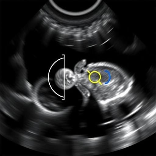
Extremely preterm infants, generally defined as those born before 28 weeks of gestation, often present with immature organ systems and are at high risk of cardiovascular instability. Close monitoring of their cardiac function is crucial, not only to detect abnormalities but also to guide therapeutic decisions in neonatal intensive care units (NICUs). Among the cardiac parameters assessed, the left atrium to aortic root ratio (LA:Ao) and fractional shortening (FS) are pivotal. The LA:Ao ratio provides a window into left atrial dilation—a marker potentially indicating increased cardiac workload or volume overload—while FS offers a quantifiable measure of left ventricular contractile function.
The study, conducted by Kanagaraj and colleagues and recently published in Pediatric Research, undertakes the significant task of comparing interrater reliability between 2D and M-mode echocardiography for measuring these critical indices in extremely preterm neonates. Interrater reliability speaks to the consistency between different observers conducting the measurements, a factor vital for ensuring diagnostic accuracy and clinical applicability. The findings shed light on the nuanced advantages and potential pitfalls inherent in these imaging modalities.
M-mode echocardiography, long favored for its temporal resolution, captures cardiac structures along a single ultrasound beam, rendering precise motion images of cardiac walls and valve function. This technique, historically regarded as the gold standard for measuring fractional shortening, excels in providing rapid, highly reproducible data in neonates whose heart rates can be alarmingly high. However, M-mode’s limitation lies in its reliance on an optimal imaging plane and precise alignment with cardiac structures, which can be technically challenging, especially in fragile neonates with small thoracic windows.
In contrast, two-dimensional echocardiography produces cross-sectional images that capture the heart’s anatomy in a planar frame, enabling concurrent visualization of multiple structures. This modality allows for a more holistic assessment, providing not only quantitative but qualitative evaluation of cardiac morphology and function. The trade-off, however, is its relative dependence on operator expertise to accurately delineate borders and measure dimensions, which may introduce variability between observers.
Kanagaraj’s study meticulously recruited a cohort of extremely preterm infants within a NICU setting. Using standardized protocols, multiple trained echocardiographers independently measured LA:Ao ratios and FS via both 2D and M-mode echocardiography. The researchers then applied statistical analyses to evaluate the interrater reliability for each measurement, using intraclass correlation coefficients (ICC) to quantify agreement levels. Their results provide insightful revelations into methodological robustness and clinical utility.
The study reports that for the LA:Ao ratio, two-dimensional echocardiography exhibited superior interrater reliability compared to M-mode. This finding underscores 2D’s advantage in capturing comprehensive anatomical relationships, allowing observers to more confidently and reproducibly identify the left atrium and aortic root boundaries. Given the clinical importance of accurately assessing left atrial dilation, with implications for fluid management and hemodynamic stability, this advantage could translate into better patient monitoring.
When addressing fractional shortening, the study found M-mode echocardiography still generally outperformed 2D in interrater agreement. This aligns with the historical precedent of M-mode’s elevated temporal resolution capturing rapid changes in ventricular dimensions during systole and diastole. However, the margin of superiority was narrower than anticipated, suggesting that with adequate training and standardized imaging protocols, 2D measurements might approach the reliability of M-mode.
Moreover, the research highlights the potential for combining both echocardiographic modalities to maximize diagnostic accuracy. In clinical practice, a dual-modality approach could harness the strength of 2D’s anatomical clarity and M-mode’s functional precision. This could empower neonatologists to detect subtle cardiovascular deviations earlier and tailor interventions appropriately.
The authors also reflect on the technical challenges inherent in echocardiographic imaging of preterm infants. Small body size, high heart rates, and variable acoustic windows necessitate meticulous technique and operator skill. They advocate for enhanced training programs focused on neonatal cardiac imaging and call for the development of consensus guidelines to harmonize measurement techniques. Such standards would reduce variability and improve the comparability of studies across institutions.
This study’s implications extend beyond neonatal cardiac imaging alone. In the realm of targeted neonatal echocardiography (TNE), which has emerged as a pivotal bedside tool, establishing reliable, reproducible measurement techniques is foundational to its broader adoption. Ensuring that different practitioners can produce consistent interpretations is essential for integrating echocardiography into routine hemodynamic assessments and therapeutic decision pathways.
Importantly, the researchers contextualize their findings within the dynamic pathophysiology of preterm neonatal hearts. The myocardium at this stage exhibits distinct contractile and compliance characteristics, and volume status rapidly fluctuates due to ongoing medical interventions such as fluid replacement, ventilation strategies, and medications. Regular, reliable echocardiographic assessment thus becomes indispensable, and tools with proven interrater reliability enhance clinical confidence.
Furthermore, the article calls attention to current gaps in knowledge, highlighting the necessity for longitudinal studies tracking cardiac function over time in extremely preterm infants. Following these parameters as infants mature could unveil new insights into the long-term cardiac sequelae of prematurity and the influence of neonatal cardiovascular management strategies.
The innovation embedded in this study lies not only in its comparative approach but also in its practical translational potential. By identifying which echocardiographic methods offer more consistent measurements, clinicians and researchers can standardize imaging approaches, improving the fidelity of cardiovascular monitoring in a particularly vulnerable population.
Additionally, advances in echocardiographic technology, such as the increasing availability of high-frequency neonatal probes and enhanced image processing algorithms, promise to further ameliorate measurement reliability. Combining these technological gains with the methodological insights from Kanagaraj’s research could revolutionize TNE practice.
In sum, this comprehensive evaluation of 2D versus M-mode echocardiography in measuring left atrium to aortic root ratio and fractional shortening in extremely preterm infants highlights nuanced strengths and limitations of each modality. It cautions practitioners about potential variability and encourages a balanced, informed choice of imaging techniques tailored to the clinical context. The study embodies a critical stride toward refining neonatal cardiac care, fostering improved outcomes through precision diagnostics.
Ultimately, as neonatal survival rates improve worldwide, the demand for sophisticated cardiovascular evaluation tools rises. Studies like this are instrumental in honing those tools, ensuring that the tiniest patients receive the most accurate insights into their cardiac health—a beacon of hope shining in the fragility of early life.
Subject of Research: The reliability of two-dimensional (2D) versus motion mode (M-mode) echocardiography for measuring left atrium to aortic root ratio (LA:Ao) and fractional shortening (FS) in extremely preterm infants.
Article Title: Reliability of two-dimensional versus M-mode echocardiography for left atrium/aortic diameter ratio and fractional shortening in extremely preterm infants.
Article References:
Kanagaraj, U.K., Castaldo, M., Braschel, M. et al. Reliability of two-dimensional versus M-mode echocardiography for left atrium/aortic diameter ratio and fractional shortening in extremely preterm infants. Pediatr Res (2025). https://doi.org/10.1038/s41390-025-04389-z
Image Credits: AI Generated
DOI: https://doi.org/10.1038/s41390-025-04389-z
Tags: cardiovascular assessment in preterm infantschallenges in assessing extremely preterm infantsechocardiography techniques for newbornsfractional shortening in preterm infantsinterrater reliability in echocardiography studiesleft atrium to aortic root ratio significanceM-mode echocardiography in neonatologyneonatal cardiac function monitoringnon-invasive cardiac diagnostics in NICUsoutcomes of extremely preterm infantsprecision medicine in neonatal carereliability of 2D echocardiography


