New research from Japanese scientists offers a glimpse of how female gametes’ cell fates are determined and shows promise for future progress in plant breeding
Credit: Issey Takahashi
Scientists from Nagoya University, Yokohama City University and Chubu University have developed a system which enables the live imaging of the formation of the female gamete in plants.
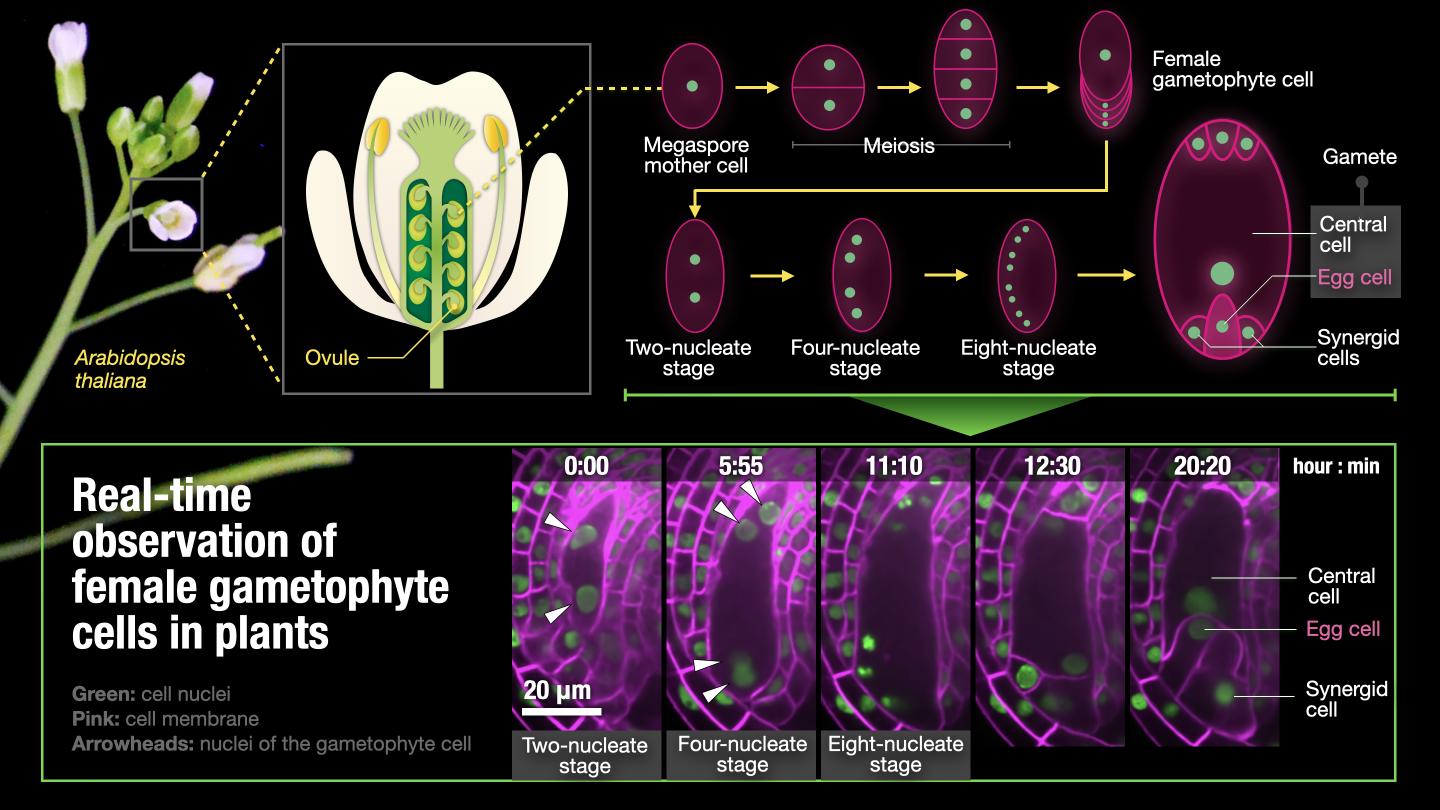
In flowering plants, the sperm cell and egg cell meet and fertilization takes place in the flower. While sperm cells are made in the pollen, egg cells are made in the ovule, the structure that becomes the seed. However, as the ovule is buried deep within the pistil, it has thus far been impossible to observe the formation of the egg cell in living plants.
The team, led by Dr Daisuke Kurihara and Dr Tetsuya Higashiyama of Nagoya University Institute of Transformative Bio-Molecules (WPI-ITbM), Dr Daichi Susaki of Yokohama City University Kihara Institute for Biological Research and Dr Takamasa Suzuki of Chubu University College of Bioscience and Biotechnology, using the ovule culturing technology that they had developed previously, succeeded in capturing images of the egg cell being formed inside the ovule. On top of that, they were able to isolate the egg cell and its neighboring cells, and by analyzing the genes expressed in these few cells, identify how the cells adjoining the egg cell determine its fate.
Living things which carry out sexual reproduction produce offspring via a fertilization process involving male and female gametes. In animals, the female gamete (the egg) is produced by meiosis, a type of cell division that halves the number of chromosomes present in the cell. However, the process in flowering plants is rather more lengthy. Following meiosis, karyomitosis (nuclear division) takes place three times within the cell, resulting in the production of a single cell with eight nuclei. This cell then divides, producing cells with a variety of different roles including two gametes, the egg cell and central cell, and the synergid cells. However, it was not yet understood precisely how the two female gametes were produced among the seven new cells that result from this process of division.
Using an ovule culturing method that they had developed previously, the research team attempted the observation in real time of the formation of the female gamete in Arabidopsis thaliana. They saw that when the first nuclear division takes place, the resulting two nuclei go to the opposite ends of the cell. Dividing again into four, the nuclei then line up along the edge of the cell. Finally, dividing again into eight, the plasma membranes are constructed around the nuclei, forming the cells which are attached to the two gametes (the egg cell and central cell). Having observed 157 cases of this division, they found that the nuclei close to where the pollen tube penetrates would become the nuclei of the synergid, egg and central cells, demonstrating that the position of the nuclei within the cell has a strong correlation with cell fate.
Continuing, in order to find out when the various cells’ fates are determined, they analyzed the time at which expression of the specific transcription factor myb98, important for the differentiation and function of the synergid cells, commenced. They found that myb98 begins to be expressed very shortly after the nuclei divide into 8 and are enclosed by the plasma membranes. Given that the specific transcription factor for the egg cell can also be found in the egg cell at the same early stage, it can be considered that cell fate is determined immediately after the formation of the plasma membranes, or possibly even earlier.
The time at which cell fate is determined is significant because it gives us an insight into how plants remain adaptable to environmental conditions by flexibly changing cell fate and thus ensuring the survival of crucial cells such as gametes.
Looking to the future, the research team’s focus will be on discovering how the cell fate change is accomplished, and explaining its molecular mechanism. Once the molecular mechanism has been analyzed, it is expected that this field of research will contribute to the development of methods to increase plant fertilization rates and environmental resistance, offering the prospect of solving key issues in food supply that affect millions of people around the world.
###
Media Contact
Daisuke Kurihara
[email protected]
Related Journal Article
http://dx.

