Pathology remains fundamental to modern clinical medicine and disease diagnosis, yet fewer than ten percent of clinical laboratories have fully transitioned from physical glass slide microscopy to digital whole-slide imaging systems. Conventional whole-slide imaging platforms present significant operational, logistical, and financial obstacles, often costing more than an entire fleet of clinical microscopes and requiring specialized personnel for scanning, quality control, and data management. Furthermore, automated scanning introduces an extra preparation step prior to sign-out without eliminating the initial glass slide production process, leaving healthcare leaders hesitant to abandon established workflows without an immediate return on investment. To bridge this persistent operational divide, researchers engineered the Histopathology Computer Assisted Microscope, known as HistoCAM, an ambient instrumentation and software platform that continuously and unobtrusively converts standard glass slide review into multi-resolution digital slide images in real time. Operating concurrently with normal examination, the system captures diagnostic sessions directly through conventional brightfield optics without modifying standard viewing behavior, imposing artificial stage speed limits, or requiring mechanical automation hardware.
Previous attempts to digitize pathology workflows or incorporate computational assistance at the microscope bench have faced substantial physical and diagnostic trade-offs. Devices such as the Augmented-Reality Microscope project artificial intelligence overlays directly into the ocular eyepieces but restrict computational evaluation and user viewing to an isolated field of view, lacking broader spatial context and retaining no memory of previously inspected tissue sections. Alternative video-mosaicking platforms relying on motorized stages or manual slide sweeping often utilize undersized image sensors that underfill the microscope field of view. Consequently, these configurations force pathologists to divert their gaze from the optical eyepieces to computer monitors to monitor stage velocity and prevent unstitched gaps, interrupting natural diagnostic routines. In contrast, HistoCAM achieves ambient digitization by passively capturing the complete eyepiece field of view across all magnification levels while the pathologist looks exclusively through the optical lenses, generating seamless multi-resolution slide images alongside rich spatiotemporal behavioral telemetry that reconstructs the entire diagnostic trajectory.
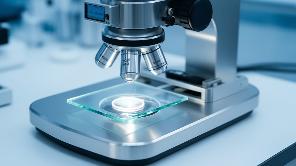
The physical architecture of HistoCAM couples standard upright brightfield microscopes to a high-speed, large-format complementary metal-oxide-semiconductor camera sensor through an optical trinocular beamsplitter. In evaluated prototype builds using Nikon Eclipse Si and Ci frames, the optical configuration directs twenty percent of the illumination to the binocular eyepieces and eighty percent to the camera sensor without intervening demagnification optics, ensuring that optical resolution is not compromised to match sensor dimensions. To avoid optical vignetting across all magnifications, a swing-out substage condenser is deployed, providing complete field-of-view coverage from low-power overview lenses to high-power objectives. The selected imaging unit, a thirty-one megapixel Sony IMX342 color sensor housed within a FLIR Oryx ten-gigabit Ethernet camera, features an APS-C format with a 27.9-millimeter diagonal, 3.45-micrometer pixels, and an active resolution of 6464 by 4852 pixels. Operating at twenty-six frames per second, this configuration yields an expansive space-bandwidth-time product of approximately 815 megapixels per second, delivering 92 to 99 percent field-of-view coverage and 98 to over 100 percent space-bandwidth product coverage across all tested microscope objectives.
Establishing the minimum operational specifications necessary to eliminate motion blur and prevent spatial gaps during unconstrained manual slide movement required extensive kinematic investigation. Using an initial high-speed prototype equipped with a FLIR Blackfly S camera recording at 200 to 250 frames per second, researchers measured the translation speeds of pathologists conducting routine glass slide examinations across different objective lenses. Hand-driven stage movements reached maximum translation speeds of seven millimeters per second with an average of 1.8 millimeters per second under a 4X objective, decreasing to a maximum of two millimeters per second and an average of 0.3 millimeters per second under a 20X objective. By modeling the relationship between sensor geometry, magnification, and stage displacement, the engineers calculated that maintaining a conservative twenty percent overlap between successive frames for reliable feature tracking required a minimum capture rate of seven frames per second, corresponding to 154.6 megapixels per second. Because the operational 31-megapixel sensor streams at twenty-six frames per second, the physical system provides substantial margin above this mechanical threshold, enabling pathologists to manipulate glass slides at unrestricted velocities without introducing gaps into the stitched mosaic.
Streaming continuous 31-megapixel Bayer raw video at high frame rates generates data volumes reaching forty-eight gigabytes per minute, creating substantial data ingestion and processing challenges. To handle this throughput in real time, the team developed a custom multi-threaded desktop software application in C++ utilizing OpenCV and JUCE libraries. Incoming raw frames are initially placed into a prioritized memory queue, debayered into RGB format using standard RGGB interpolation patterns, and subjected to automated quality control algorithms evaluating blur and illumination. Frames failing exposure or focus thresholds, along with three adjacent frames preceding and following them to catch transition artifacts during objective nosepiece rotation, are discarded. For accepted frames, cropped and downsampled representations are processed on the host central processing unit using Oriented FAST and Rotated BRIEF, known as ORB, to compute single-scale feature descriptors. Pairwise feature matching chains adjacent frames together sequentially to establish absolute spatial coordinates, transferring full-resolution image buffers to graphic processing unit memory only when validated for compositing, thereby maintaining strict system memory constraints.
To assemble high-resolution mosaics without computational bottlenecks, the software implements an approximate graph-based compositing algorithm that treats frame selection as a minimum-set coverage problem. Because finding an optimal subset of overlapping frames is mathematically NP-hard, the platform models incoming registered frames as geometric sites within a dynamic Voronoi diagram. The system evaluates the polygonal Voronoi face assigned to each newly positioned frame, which delineates the spatial region where that frame contributes optical data closer to its lens center than any neighboring frame, taking advantage of superior optical quality near the central axis. If the calculated area of the contributing Voronoi face encompasses less than twenty percent of the total frame surface, the frame is rejected as redundant and removed from the topological graph. Accepted frames contribute their bounded Voronoi face as a copy-mask, projecting updated pixels onto designated composite image tiles. This geometric approach naturally accommodates static pauses when a clinician steps away from the microscope; static duplicate frames yield negligible Voronoi face contributions and are immediately purged, preventing memory backlog and data pileup.
Accommodating the non-linear manner in which pathologists switch magnifications required an automated, scale-aware image registration pipeline capable of handling dramatic jumps in resolution. Because single-scale ORB descriptors fail across differing optical magnifications, incoming frames collected after an objective lens rotation fail to register against prior components. When an accepted frame fails pairwise matching against existing single-scale coordinates, the system designates it as a root frame, initiating a new connected component in the image hierarchy. The software extracts Scale Invariant Feature Transform, or SIFT, keypoints from the root image and executes multi-resolution GPU matching against constituent frames of earlier components. A single successful cross-scale SIFT match establishes the scale factor and spatial offset necessary to position the new component relative to the global mosaic origin. New image layers are indexed into a tiled pyramidal hierarchy where higher-resolution data consistently takes rendering precedence, allowing instantaneous multi-resolution slide image generation spanning 2X, 4X, 10X, 20X, and 40X magnifications during natural diagnostic review.
Because incremental pairwise coordinate chaining can accumulate spatial drift and produce visible stitching seam misalignments at closed inspection loops, HistoCAM integrates an optional GPU-accelerated bundle adjustment framework. Across standard continuous acquisition sessions, positional deviation between accumulated coordinates and direct pairwise homographies averaged approximately eighteen pixels, rarely exceeding eighty pixels. Standard global bundle adjustment algorithms often introduce severe numerical instability when optimizing multi-scale microscopy frames simultaneously, largely because unconstrained rotation parameters around three axes introduce non-linear complexity unnecessary for planar slide microscopy. To resolve this, HistoCAM executes bundle adjustment independently within each single-magnification component before treating each aligned component as a unified planar element in a secondary cross-scale optimization. This hierarchical bundle adjustment reduces maximum alignment error to nine pixels across experimental benchmarks, processing a composite containing 215 frames across four optical objectives in approximately eight seconds by overlapping frame retrieval with mathematical system setup.
Beyond assembling composite imagery, HistoCAM introduces ambient datafication by recording continuous spatiotemporal behavioral telemetry of clinical slide examination. By coupling frame timestamps to spatial coordinates and objective turret states, the platform logs exactly where a practitioner looked, the sequence of tissue exploration, and the precise dwell time spent inspecting individual microscopic regions. These interaction data allow the automated generation of magnification utilization maps, dwell-time heatmaps, and chronologically encoded navigational vector paths across the tissue specimen. Unlike digital whole-slide viewing software where viewport coordinates captured via computer mouse movements frequently incorporate noisy, unintentional cursor drift, microscope-based physical navigation reflects deliberate mechanical actions, such as manual stage adjustments and physical objective lens selections. These telemetry layers can support real-time quality assurance by alerting pathologists to uninspected tissue margins or providing immediate dwell-time saliency checks, while also enabling researchers to compare glass slide diagnostic behaviors directly against digital viewing workflows.
The clinical utility of HistoCAM was demonstrated through structured workflow uplifts during the evaluation of complex surgical pathology specimens, such as radical prostatectomy resections. In routine clinical practice, evaluating prostatectomy cases requires examining between 50 and 150 glass slides per patient to assess International Society of Urological Pathology Gleason Grade Groups, quantify overall tumor volume, and measure the linear extent of positive surgical margins. Traditionally, pathologists outline suspicious areas manually using ink pens directly on the glass slide surfaces, afterward estimating tumor burden and margin distances through visual eyeballing, calipers, or handheld plastic rulers. This conventional analog process is semi-quantitative and represents a documented source of inter-observer diagnostic variability. Utilizing the HistoCAM application, pathologists can immediately delineate pathological lesions on the real-time composite using digital polygon annotation tools. Because the physical pixel scale is mathematically calibrated across all magnifications, the platform automatically calculates precise tumor surface areas, computes the exact percentage of tumor involvement relative to the entire specimen boundary, and measures positive surgical margin lengths, storing auditable digital records without disrupting microscopic sign-out.
The platform also incorporates native real-time artificial intelligence inference capabilities operating at both patch and whole-slide levels. As pyramidal image tiles are updated during compositing, background processes batch and submit them to pre-configured deep learning encoders. For tile-level detection tasks where spatial context from surrounding areas is not required, embeddings are classified directly using lightweight multilayer perceptrons. In an experimental demonstration detecting acid-fast bacilli within sputum smear preparations, a PathDINO foundation model accurately flagged suspected bacterial clusters at 4X and 10X magnifications, guiding the microscopist to center the field of view and confirm bacterial presence under a 40X objective. For contextual whole-slide applications, such as Gleason grading on prostatectomy sections, the system passes continuous tile embeddings generated by a Prov-Gigapath DINOv2 vision encoder into an attention-based slide aggregator, classifying contextualized token representations to grade individual tiles according to glandular architecture. Additionally, integration of Segment Anything Model 2 allows clinicians to produce detailed regional tissue annotations rapidly using interactive prompt clicks, automatically partitioning segmented masks into pre-embedded tile collections suitable for training future computational pathology models.
To facilitate clinical deployment in resource-constrained environments that cannot accommodate complex multi-GPU computer chassis, the HistoCAM architecture was transferred from an initial dual-GPU AMD Ryzen Threadripper workstation to a compact NVIDIA DGX Spark edge computing system. Featuring 128 gigabytes of unified system memory shared between the central processor and graphics hardware, the unified edge system avoids data bus transfer bottlenecks between discrete memory banks, executing image ingestion, Voronoi compositing, and neural inference within a unified memory space. Crucially, because HistoCAM selectively captures only those regions actively inspected by the clinician at high magnification while preserving lower-resolution overviews elsewhere, the resulting multi-resolution slide images are highly data-efficient. The raw file sizes of HistoCAM composites average between five and ten percent of uniform 20X whole-slide scans, rendering them forty to eighty times smaller than uniform 40X digital files. By coupling compact edge hardware with targeted, behaviorally driven data capture, HistoCAM provides an accessible bridge from traditional analog microscopy to process-aware computational pathology.
Subject of Research: Real-time ambient digitization and behavioral datafication of glass slide microscopy
Article Title: Ambient, real-time digitization and datafication of glass slide microscopy towards AI-at-the-microscope
Article References: Maira, C., Cooper, M. S., Ashman, K. L., Sholl, A. B., Fox, S. E., Halat, S., Manthey, D., Choudhury, R., Sears, J., Wenk, C., Brown, J. Q., & Summa, B. (2026). Ambient, real-time digitization and datafication of glass slide microscopy towards AI-at-the-microscope. Nature Communications, 17(1), Article 10933. https://doi.org/10.1038/s41467-026-77887-1
Image Credits: AI Generated
DOI: 10.1038/s41467-026-77887-1
Keywords: HistoCAM, Digital Pathology, Brightfield Microscopy, Multi-Resolution Slide Images, Space-Bandwidth-Time Product, Voronoi Compositing, Computational Pathology, Whole-Slide Imaging, Diagnostic Telemetry, Bundle Adjustment, PathDINO, Prov-Gigapath, Segment Anything Model 2, Gleason Grading, Acid-Fast Bacilli, Unified Memory Computing, Edge AI
News Source: Ophelia Keating. (November 16, 2026). Real-Time Microscope Platform Converts Routine Glass Slide Pathology into Multi-Resolution Digital Data. Scienmag.