Clinicians have documented the rare endoscopic removal and pathological confirmation of an intraluminal polypoid rectal lesion composed of ectopic cutaneous, subcutaneous, and thyroid tissues in an elderly patient. The report, published in BMC Gastroenterology, describes the diagnosis and complete excision of this composite heterotopic growth, which presented an unusual combination of tissue types within the lower gastrointestinal tract.
Heterotopic thyroid tissue represents an uncommon congenital anomaly that arises from the aberrant embryological migration of the thyroid primordium. While the majority of such heterotopic lesions develop along the thyroglossal duct within the head-and-neck region, their occurrence as an intraluminal rectal lesion is exceedingly rare in the medical literature. The simultaneous presence of heterotopic thyroid tissue alongside ectopic skin and subcutaneous adipose layers in the gastrointestinal tract introduces significant diagnostic challenges for evaluating physicians and pathologists.
The clinical case involved a 79-year-old female who was admitted to an emergency department following four hours of fresh bright-red hematochezia. Prior to the episode, the patient had no documented medical history of thyroid disease or gastrointestinal malignancy, presenting with acute bleeding as the primary clinical manifestation.
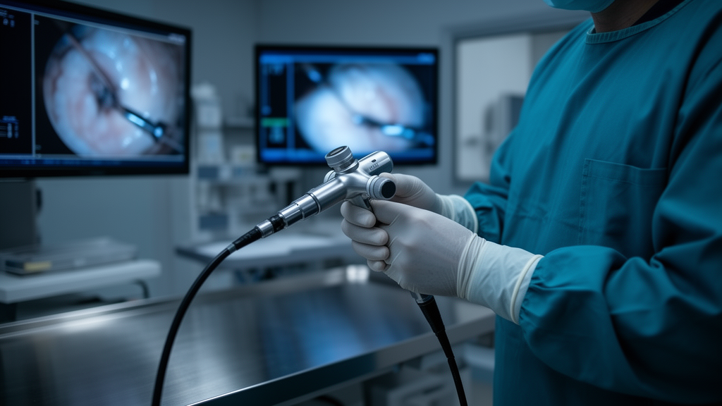
During an endoscopic examination, colonoscopy revealed a solitary, pale-coloured polypoid lesion with a smooth mucosal surface and a short pedicle. Situated approximately 10 centimetres from the anal verge and measuring roughly 1.5 centimetres in diameter, the lesion was completely excised using endoscopic mucosal resection without procedural complications.
Subsequent histopathological evaluation revealed that the resected specimen consisted of cutaneous stratified squamous epithelium and subcutaneous adipose tissue situated above nests of benign-appearing thyroid follicles. Immunohistochemical testing confirmed the thyroid lineage of the ectopic follicles through strong positive staining for thyroid transcription factor-1, thyroglobulin, and PAX-8, accompanied by focal cytokeratin positivity and negative CD10 expression, with no signs of cellular atypia or malignancy.
Following the intervention, the patient experienced an uneventful recovery and remained asymptomatic during subsequent follow-up. The authors concluded that the smooth, pale endoscopic appearance provides a subtle auxiliary clue for identification, while complete endoscopic resection represents a feasible intervention for benign composite lesions, supported by individualized long-term surveillance to monitor for potential recurrence or tissue transformation.
Subject of Research: Heterotopic thyroid and cutaneous tissue presenting as a polypoid rectal lesion
Article Title: Polypoid rectal lesion composed of ectopic cutaneous, subcutaneous, and thyroid tissues: a case report
Article References: Kou, J., Li, S., & Chen, Y. (2026). Polypoid rectal lesion composed of ectopic cutaneous, subcutaneous, and thyroid tissues: a case report. BMC Gastroenterology. https://doi.org/10.1186/s12876-026-05455-4
Image Credits: AI Generated
DOI: 10.1186/s12876-026-05455-4
Keywords: Heterotopic thyroid tissue, Ectopic cutaneous tissue, Subcutaneous adipose tissue, Polypoid rectal lesion, Endoscopic mucosal resection, Hematochezia, Thyroid transcription factor-1, Thyroglobulin, PAX-8, Colonoscopy, Gastrointestinal tract, Histopathology, Immunohistochemical staining, Thyroid primordium, Thyroglossal duct, Cytokeratin, CD10
News Source: Ophelia Keating. (November 12, 2026). Rare Rectal Polyp Containing Ectopic Thyroid and Skin Tissues Removed Endoscopically. Scienmag.