• HOME
  • NEWS
  • EXPLORE
    • CAREER
      • Companies
      • Jobs
    • EVENTS
    • iGEM
      • News
      • Team
    • PHOTOS
    • VIDEO
    • WIKI
  • BLOG
  • COMMUNITY
    • FACEBOOK
    • INSTAGRAM
    • TWITTER
Saturday, November 1, 2025
BIOENGINEER.ORG
No Result
View All Result
  • Login
  • HOME
  • NEWS
  • EXPLORE
    • CAREER
      • Companies
      • Jobs
        • Lecturer
        • PhD Studentship
        • Postdoc
        • Research Assistant
    • EVENTS
    • iGEM
      • News
      • Team
    • PHOTOS
    • VIDEO
    • WIKI
  • BLOG
  • COMMUNITY
    • FACEBOOK
    • INSTAGRAM
    • TWITTER
No Result
View All Result
Bioengineer.org
No Result
View All Result
Home NEWS Science News Biology

Queen Mary University of London acquires first Cell DIVE multiplexed imager for a university outside of North America

Bioengineer by Bioengineer
November 30, 2022
in Biology
Reading Time: 2 mins read
0
An example of the Cell DIVE's insight into cells
Share on FacebookShare on TwitterShare on LinkedinShare on RedditShare on Telegram

Queen Mary University of London has become the first university outside of North America to acquire a Cell DIVE imager, providing access to the latest technology and ensuring that we remain world-leading in the fast-moving area of single-cell approaches.

An example of the Cell DIVE's insight into cells

Credit: Queen Mary University of London/Dr Adrian Biddle

Queen Mary University of London has become the first university outside of North America to acquire a Cell DIVE imager, providing access to the latest technology and ensuring that we remain world-leading in the fast-moving area of single-cell approaches.

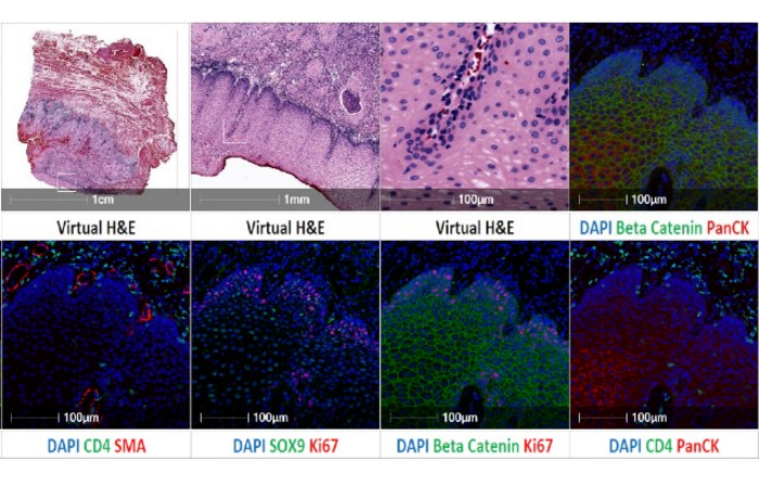
The Cell DIVE provides multiplexed imaging of tissue sections enables high-dimensional tissue imaging for quantitative analysis of at least 60 biomarkers, visualising the inner workings of single cells on a level that was previously unattainable.

What makes this technology platform significant is the integration of biology, precision imaging, sophisticated analysis software to enable deep insights at the single cell level. This offers a powerful approach to understanding the molecular and cellular processes driving tissue pathology in human disease.

The Cell DIVE is the latest addition to the Phenotypic Screening Facility at Queen Mary which is open to all staff across the University and external users and is fully supported by dedicated staff.

Prof. Cleo Bishop, Director of the Queen Mary University of London Phenotypic Screening Facility, said: “We’re excited to have this new technology available for use at the University following a successful Wellcome Trust multi-user equipment grant. The equipment will help us further our understanding of health tissues and heterogeneity within disease.”

“In the past researchers have been able to study how two to three different proteins relate to one another, but the Cell DIVE enables us to visualise more than 60 different proteins on a ‘cell-by-cell’ basis. We can directly relate each of these proteins to one and another within an individual cell, between cells and within the tissue microenvironment. This powerful approach can be used to shed new light on any area of medical research.”

As part of its 2030 strategy, the University is providing a world-class virtual and physical research infrastructure, which is available to all researchers, supported by internal and significant external funding to support academics to pioneer research and innovation.

Queen Mary has been ranked joint seventh in the UK for the quality of its research, according to the Research Excellence Framework 2021; and across the University, 92 per cent of Queen Mary’s research has been assessed as internationally excellent or world-leading according to the REF21 results.


Subject of Research

Cells

Share12Tweet7Share2ShareShareShare1

Related Posts

Decoding Boeremia exigua: Fungal Pathogen of Ginseng

Decoding Boeremia exigua: Fungal Pathogen of Ginseng

November 1, 2025
blank

Alveolar Macrophages Predict TST/IGRA Conversion Resistance

November 1, 2025

Intestinal Parasites in Punjab’s Rock Pigeons Unveiled

November 1, 2025

Rj4 Immunity Network Limits Soybean-Rhizobia Symbiosis

November 1, 2025

POPULAR NEWS

  • Sperm MicroRNAs: Crucial Mediators of Paternal Exercise Capacity Transmission

    1295 shares
    Share 517 Tweet 323
  • Stinkbug Leg Organ Hosts Symbiotic Fungi That Protect Eggs from Parasitic Wasps

    312 shares
    Share 125 Tweet 78
  • ESMO 2025: mRNA COVID Vaccines Enhance Efficacy of Cancer Immunotherapy

    203 shares
    Share 81 Tweet 51
  • New Study Suggests ALS and MS May Stem from Common Environmental Factor

    137 shares
    Share 55 Tweet 34
/div>

About

We bring you the latest biotechnology news from best research centers and universities around the world. Check our website.

Follow us

Recent News

Decoding Boeremia exigua: Fungal Pathogen of Ginseng

Manganese Catalysis: Alkylation of Arenes via Alcohols

CircSipa1l1 Drives Melanoma Differentiation via IGF2BP1 Pathway

Subscribe to Blog via Email

Enter your email address to subscribe to this blog and receive notifications of new posts by email.

Join 67 other subscribers
  • Contact Us

Bioengineer.org © Copyright 2023 All Rights Reserved.

Welcome Back!

Login to your account below

Forgotten Password?

Retrieve your password

Please enter your username or email address to reset your password.

Log In
No Result
View All Result
  • Homepages
    • Home Page 1
    • Home Page 2
  • News
  • National
  • Business
  • Health
  • Lifestyle
  • Science

Bioengineer.org © Copyright 2023 All Rights Reserved.