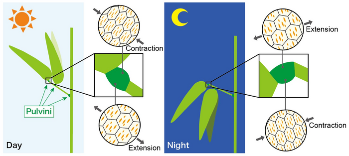
Ikoma, Japan – Plant movement has been long fascinated by many researchers. Legumes are a group of plants famous for exhibiting various leaf movements, including “nyctinastic movement”, in which the leaves open in the day and close at night. Similar plant movements include blue light-induced and touch-sensitive movements, such as in sensitive plants like Mimosa pudica. Movement in leaf structures is caused by repeated and reversible extension and contraction of “motor cells”, which are the cells in a structure called the “pulvinus” at the base of the leaflets and petioles. Such repetitive and reversible cell extension and contraction are very rare in plant cells, which are surrounded by a rigid cell wall. Moreover, it is not well understood how motor cells are capable of repetitive and reversible extension and contraction.

Credit: Miyuki T Nakata
Ikoma, Japan – Plant movement has been long fascinated by many researchers. Legumes are a group of plants famous for exhibiting various leaf movements, including “nyctinastic movement”, in which the leaves open in the day and close at night. Similar plant movements include blue light-induced and touch-sensitive movements, such as in sensitive plants like Mimosa pudica. Movement in leaf structures is caused by repeated and reversible extension and contraction of “motor cells”, which are the cells in a structure called the “pulvinus” at the base of the leaflets and petioles. Such repetitive and reversible cell extension and contraction are very rare in plant cells, which are surrounded by a rigid cell wall. Moreover, it is not well understood how motor cells are capable of repetitive and reversible extension and contraction.
Plant cell walls are composed of a number of cellulose microfibrils that shrink or expand in response to osmotic concentration differences between the inside and outside of the cell. However, the amount of change that can be induced by anisotropy in the arrangement of cellulose microfibrils cannot explain the full range of movement of the pulvinus.
A research team led by Miyuki Nakata and Taku Demura at the Nara Institute of Science and Technology (NAIST) examined the cross-sections of pulvinar motor cells from Desmodium paniculatum using confocal laser microscopy to investigate the mechanism of repetitive and reversible cell extension and contraction. They identified unique circumferential “slits” in the cell wall of the motor cells that contained less cellulose. The structures were conserved across two subfamilies of legumes, including soybeans, kudzu and sensitive plants.
Upon transferring tissue slices from legume cortical motor cells to solutions of different osmolarity, the pulvinar slits increased in width, indicating a mechanism by which plant cell walls could flex in response to solutions of different osmolarity.
Through a combination of detailed cell wall analysis, computer simulations, and observations of pulvinar slits in cells undergoing extension and contraction, pulvinar slits were determined to be mechanically flexible structures that open and close during cell extension and contraction. “Computer modeling suggested that pulvinar slits facilitate anisotropic extension in the direction perpendicular to the slits in the presence of turgor pressure,” says Miyuki Nakata. The researchers compared the action to the straight cuts or slits used in kirigami, a Japanese papercraft, to enhance the extensibility of the paper sheet.
Thus, the research team proposed that these unique, pulvinar slits are structures that act to allow more movement of the cortical motor cells than would otherwise be allowed by the typical cellulose microfibrils in the cell wall.
“We provide a hypothesis that pulvinar slits have a role in dynamic leaf movement through repetitive and reversible deformation of cortical motor cells in concert with other factors including cellulose orientation, pectin-rich composition of the cell wall, the geometry of cortical motor cells, and the actin cytoskeleton,” says Miyuki Nakata.
###
Resource
Title: Pulvinar slits: Cellulose-deficient and de-methyl-esterified pectin-rich structures in a legume motor cell
Authors: Masahiro Takahara, Satoru Tsugawa, Shingo Sakamoto, Taku Demura, Miyuki T Nakata
Journal: Plant Physiology
Information about the Plant Metabolic Regulation Laboratory can be found at the following website: https://bsw3.naist.jp/eng/courses/courses104.html
Journal
PLANT PHYSIOLOGY
DOI
10.1093/plphys/kiad105
Article Title
Pulvinar slits: Cellulose-deficient and de-methyl-esterified pectin-rich structures in a legume motor cell


