In an analytical review published in Discover Nano, researchers have systematically categorized how self-assembled DNA nanostructures and scaffolded DNA origami serve as programmable nanoscale architectures to regulate and enhance fluorescence-based biosensing. Biological target detection and quantification underpin clinical diagnostics, environmental monitoring, and biomedical research, yet conventional fluorescence biosensors frequently face constraints such as limited sensitivity, low specificity, and operational degradation when deployed in complex biological environments. Over four decades of development, structural DNA nanotechnology has addressed these challenges by exploiting the predictable Watson–Crick base pairing of nucleic acid sequences to construct geometric frameworks with nanometer-scale addressability. By organizing functional elements such as fluorophores, quenchers, aptamers, enzymes, and metallic nanoparticles at defined coordinates, these architectures transform passive DNA scaffolding into active signal-transduction platforms capable of generating amplified, quantitative, and multiplexed optical readouts across diverse bioanalytical settings.
The underlying mechanics of these platforms rely on fundamental photophysical signal transduction pathways, primarily Förster resonance energy transfer, nanosurface energy transfer, photoinduced electron transfer, collisional quenching, de novo fluorophore generation, and plasmon-enhanced fluorescence. Förster resonance energy transfer operates via non-radiative dipole–dipole coupling between an excited donor fluorophore and an acceptor chromophore, exhibiting an efficiency that decreases with the sixth power of intermolecular separation and offering extreme sensitivity to distance shifts between two and ten nanometers. This distance dependency enables ratiometric fluorescence sensing, where dual emission channels normalize internal variations in probe concentration, excitation intensity, and fluctuating test environments. Alternatively, turn-on and turn-off sensing mechanisms modulate proximity between fluorophores and quenchers, while de novo pathways generate emitters only following molecular recognition, such as synthesizing silver nanoclusters on cytosine-rich sequences, assembling copper nanoparticles on poly-thymine templates, or binding fluorogenic dyes to split RNA aptamers like Broccoli and Spinach.
Among non-origami architectures, three-way junction DNA nanostructures provide an independently programmable three-branched geometry that acts as a localized reaction hub. Early iterations utilized junction assembly and disassembly for label-free detection, such as monitoring the binding of tobramycin using aptamer branches where junction dissociation decreased double-stranded regions accessible to intercalating PicoGreen dye. Subsequent designs utilized proximity-localized catalytic hairpin assembly, bringing hairpin probes into nanoconfined proximity to accelerate reaction kinetics without solution-phase diffusion bottlenecks, ultimately directing the synthesis of fluorescent silver nanoclusters. Three-way junctions have also organized multi-cluster arrays of silver nanoclusters for ratiometric nucleic acid quantification and directed split Broccoli aptamer reconstruction for Campylobacter DNA detection at femtomolar concentrations. Furthermore, integrating three-way junctions with terminal deoxynucleotidyl transferase and Nt.BspQI nicking endonuclease cascade amplification produced poly-thymine templates for fluorescent copper nanoparticle synthesis, achieving a one-femtomolar detection limit for microRNA-153-3p while differentiating expression levels in clinical blood samples from Parkinson’s disease patients and healthy individuals.
Hyperbranched and dendritic DNA nanostructures offer significantly expanded surface areas and high probe-loading capacities, enabling single target-recognition events to trigger multiple fluorophore assemblies. Synthesized via repetitive enzymatic ligation or through amplification mechanisms including rolling circle amplification and hybridization chain reaction, these branched frameworks create nanoconfined reaction volumes that enhance hybridization efficiency. Illustrative platforms combine double-stranded branch migration, hybridization chain reactions, and DNAzyme feedback circuits for the sensitive quantification of Pseudomonas aeruginosa. In other configurations, hyperbranched rolling circle amplification architectures provide dense binding matrices for ratiometric fluorescence probes, mitigating environmental measurement errors. When combined with CRISPR-Cas12a systems and the amplification refractory mutation system on gold nanomagnetic beads, dendritic nanotrees present abundant cleaved reporters to amplify signals for the FecB single-nucleotide mutation. Additionally, hyperbranched networks templating dense silver nanoclusters have been coupled with metal-aggregation-induced emission luminogen frameworks, enabling dual-emission ratiometric Förster resonance energy transfer sensing for the environmental pollutant 17beta-estradiol.
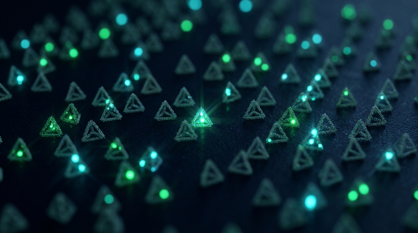
Tetrahedral DNA nanostructures, assembled from four single-stranded DNA oligonucleotides into rigid pyramidal geometries, present functional probes in upright, oriented configurations that alleviate steric hindrance. By modifying individual edges or vertices, researchers have realized multiplexed sensing platforms capable of simultaneously detecting distinct microRNAs, including miR-21, miR-221, and miR-155, or multiplexing enzyme and protein targets such as endonucleases, vascular endothelial growth factor, adenosine triphosphate, and thrombin without cross-interference. When configured with hairpin probes at tetrahedral vertices, localized catalytic hairpin assembly accelerates reaction cascades, as demonstrated in CRISPR-Cas12a-assisted aflatoxin B1 detection generating fluorescent G-quadruplex crystal violet complexes, and living-cell imaging of miR-21 with a detection limit of 1.32 femtomolar. Clinical utility has been shown in colorectal cancer serum panels, where tetrahedral probes maintained stability in ten percent human serum, achieving sensitive discrimination. Tetrahedral platforms have similarly enhanced uracil DNA glycosylase activity detection down to 8.6 times 10 to the minus five units per milliliter and improved Cas12a trans-cleavage kinetics against methicillin-resistant Staphylococcus aureus to eighteen colony-forming units per milliliter.
Three-dimensional DNA hydrogels formed via complementary hybridization, self-assembly, or enzymatic amplification provide porous matrices that combine target enrichment, nanoconfined catalytic reactions, and signal transduction. These networks manipulate the local microenvironments of embedded fluorophores, notably activating aggregation-induced emission luminogens by restricting their non-radiative intramolecular rotational and vibrational motions. Target-induced modulation of hydrogel assembly has been successfully applied to prostate-specific antigen quantification in patient-derived serum and label-free DNA detection using water-soluble natural luminogens. When functionalized with cytosine-rich sequences, hydrogels template uniform silver nanoclusters for microRNA quantification, while quantum dot-embedded hydrogels responsive to CRISPR-Cas13a trans-cleavage drive toehold-mediated strand displacement cascades to quench quantum dot emission. Structurally integrated platforms have further incorporated hydrogels into three-dimensionally printed microextractors, using strand displacement amplification to contract the hydrogel network and aggregate tetraphenylethene fluorophores for direct detection of free antibiotic resistance genes in environmental wastewater, aquaculture water, and fish samples.
Other spatially organized geometries, such as rolling circle amplification-derived DNA nanoflowers, spherical DNA frameworks, and constrained circular DNAzymes, provide multifunctional scaffolds capable of encapsulating molecular detection machinery. DNA nanoflowers synthesize dense cadmium-telluride quantum dots in situ or package CRISPR-Cas12a complexes alongside circular fluorescent reporters, as demonstrated by the InCasor platform for direct intracellular and ex vivo imaging of mitochondrial DNA mutations in tumor-bearing mice. Renal-clearable CRISPR nanosensors similarly enable non-invasive longitudinal urine monitoring of tumor progression and lung metastasis in living models. Self-assembled spherical DNA frameworks provide nanoreactor chambers that concentrate Cas12a-crRNA complexes and catalytic hairpin assembly reporters, facilitating H1N1 viral detection across chicken serum, bovine serum, and milk matrices. Furthermore, re-engineering linear DNAzymes into topologically rigid circular architectures regulates catalytic reaction rates, generating temporally encoded fluorescence outputs that discriminate multiple analytical targets.
Dynamic DNA nanostructures transition biosensing from static scaffolding to active molecular machines by converting target recognition into mechanical motion, structural reconfiguration, or continuous locomotion. Three-dimensional DNA walkers migrate along predefined tracks on particle surfaces or nanostructured interfaces, driving successive strand displacement and nicking endonuclease reactions that activate multiple fluorescent reporters per single recognition trigger. Localized three-dimensional walkers have enabled rapid isothermal detection of Zika virus-related nucleic acids and deoxynivalenol mycotoxin, while single-legged walkers on gold nanoparticles detect prokaryotic pathogens in extracted bacterial genomes. Advanced dynamic circuits further couple catalytic hairpin assembly, RNA-cleaving DNAzyme junctions, and circular CRISPR RNA switches to drive repetitive walker migration for detecting atherosclerosis-associated microRNAs. Similarly, particle-interacting walkers utilizing DNAzyme-functionalized walking particles repeatedly cleave fluorophores on substrate beads, substantially magnifying signal amplification efficiency for human immunodeficiency virus DNA detection.
Scaffolded DNA origami provides exceptional architectural complexity, folding a long single-stranded DNA scaffold via hundreds of staple strands to position functional molecules with sub-nanometer precision across hundreds of addressable loci. Within plasmon-enhanced fluorescence sensing, DNA origami arranges metallic nanostructures and fluorophores at defined spatial intervals, optimizing localized surface plasmon resonance while preventing contact-induced metallic quenching. The open-hotspot NACHOS nanoantenna architecture achieves up to 461-fold fluorescence enhancement, enabling smartphone-based single-molecule detection in human blood serum, while nanopatterned origami structures achieve ten-attomolar digital single-molecule DNA detection in human blood plasma. Plasmonic nanoantenna designs have expanded to gold-silver nanostar dimers for single-molecule adenosine triphosphate sensing and rhodium nanocube dimers that enhance deep-ultraviolet autofluorescence from protein tryptophan residues, realizing label-free single-molecule streptavidin detection.
Beyond plasmonic amplification, DNA origami platforms govern distance-dependent Förster resonance energy transfer and non-FRET quenching configurations with exceptional geometric fidelity. Dynamic origami hinges, levers, and books convert target-induced conformational alterations and strand displacement into quantitative fluorescence shifts. Chemically modified origami books featuring locked nucleic acids and 2-prime-fluoro modifications enable direct quantification of circulating microRNAs in clinical plasma without enzymatic amplification, while reconfigurable rotating origami nanodevices permit regenerable target detection via reversible strand exchange. Membrane-anchored origami architectures function as biophysical probes, quantifying membrane potential, surface charge, and vesicle curvature between 50 and 300 nanometers. In non-FRET configurations, origami localizes catalytic hairpin circuits on its surface for intracellular microRNA imaging, aligns aptamers to enhance sandwich immunoassay efficiency, monitors cell-surface receptor binding in real time, and drives mechanical gripper reconfigurations, exemplified by the photonic crystal-coupled DNA NanoGripper detecting SARS-CoV-2 in clinical saliva.
Despite substantial bioanalytical advancements across biological matrices, several critical technical limitations hinder the clinical translation of DNA nanostructure-based fluorescence biosensors. Physiological environments subject unprotected DNA architectures to endonuclease degradation, cation depletion, and pH fluctuations, destabilizing dynamic assemblies. Although protective polymer coatings and silica encapsulation enhance biostability, they often restrict target accessibility and hinder functional site addressability. Furthermore, manufacturing bottlenecks—including the prohibitive cost of chemically modified staple oligonucleotides, modest origami folding yields, and the scarcity of standardized large-scale fabrication protocols—restrict commercial scalability. Assembly batch-to-batch variability and structural non-uniformity introduce analytical signal discrepancies, particularly in plasmon-enhanced systems highly sensitive to interparticle spacing. Conventional organic fluorophores also remain vulnerable to photobleaching and optical blinking, motivating shifts toward quantum dots, upconversion nanoparticles, and metal nanoclusters. Addressing matrix-induced background interference, streamlining sample preparation, and integrating programmable architectures with microfluidics, portable detection hardware, and artificial intelligence-assisted image analysis remain necessary requirements for real-world clinical diagnostic implementation.
Subject of Research: Fluorescence biosensing platforms enabled by structural DNA nanotechnology and DNA origami
Article Title: Fluorescence-based biosensing with DNA nanostructures and DNA origami: from programmable architectures to quantitative readouts
Article References: Kim, S., Choi, M. Y., Jeong, H., Ahn, H. R., & Choi, J.-H. (2026). Fluorescence-based biosensing with DNA nanostructures and DNA origami: from programmable architectures to quantitative readouts. Discover Nano, 21(1), Article 603. https://doi.org/10.1186/s11671-026-05083-7
Image Credits: AI Generated
DOI: 10.1186/s11671-026-05083-7
Keywords: DNA nanotechnology, DNA origami, fluorescence biosensing, FRET, plasmon-enhanced fluorescence, tetrahedral DNA nanostructures, CRISPR biosensing, molecular diagnostics, Fluorescence-based, biosensing, nanostructures, origami
News Source: Denise Maddox. (November 7, 2026). Programmable DNA Nanostructures Advance Quantitative Fluorescence Biosensing Across Biological Diagnostics. Scienmag.