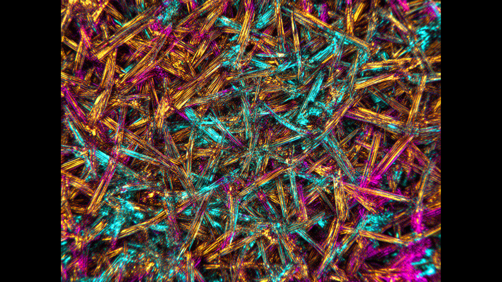
Quantitative polarized light microscopy has revealed significant reductions in optical retardance across disrupted collagen layers of mouse tympanic membranes affected by chronic otitis media. In the study conducted by researchers at Rosalind Franklin University of Medicine and Science and Northwestern University, structurally altered regions of diseased membranes exhibited a mean optical retardance of 4.62 ± 1.34 nm, reflecting localized degradation and disorganization of the underlying tissue architecture.
Chronic otitis media causes progressive structural remodeling within the tympanic membrane, initiating with epithelial hyperplasia before advancing to the disruption of the organized collagen framework responsible for its mechanical behavior. This deterioration of collagen integrity is clinically associated with an increased susceptibility to membrane retraction and perforation. However, clinicians currently lack an established method capable of directly evaluating layer-specific tympanic membrane microstructure in vivo.
To investigate microstructural changes during disease progression, the investigators intranasally inoculated mice to induce otitis media. Middle ear involvement was monitored and confirmed via auditory brainstem response measurements recorded at baseline prior to inoculation, as well as at 7 and 28 days post-inoculation.
At 28 days post-inoculation, tympanic membranes from both control mice and infected mice were harvested, embedded, and sectioned at a thickness of 7 µm. The team then quantified optical retardance in the collagen layers using quantitative polarized light microscopy to assess the extent of structural alteration.
Microscopic measurements demonstrated distinct regional differences in tissue organization at 28 days post-inoculation. While disrupted membrane regions exhibited a mean retardance of 4.62 ± 1.34 nm, adjacent unaltered regions within the very same tympanic membranes showed a significantly higher mean retardance of 9.78 ± 0.95 nm. Healthy control tympanic membranes displayed comparable values of 9.16 ± 0.87 nm, with retardance in altered regions measuring significantly lower than both unaffected control tissue and adjacent unaltered zones (p < 0.05 for both comparisons).
The utility of polarized light microscopy for identifying collagen breakdown was also validated in harvested human tympanic membranes subjected to experimental heat damage, which produced retardance changes similar to those recorded in the mouse model. The authors noted that establishing quantitative polarized light microscopy metrics offers biological support for polarization-sensitive optical coherence tomography, an imaging modality that could eventually be integrated into specialized otoscopes to assess tympanic membrane layer integrity in living patients.
Subject of Research: Microstructural collagen alterations in tympanic membranes during chronic otitis media
Article Title: Polarized light microscopy to quantify changes in the tympanic membrane microstructure in chronic otitis media
Article References: Villa, J., Cobas, D., Cury, J., Rand, D., Tan, X., Reynolds, J., Ahmed, Z., Stutzmann, G. E., & Richter, C.-P. (2026). Polarized light microscopy to quantify changes in the tympanic membrane microstructure in chronic otitis media. Frontiers in Audiology and Otology, 4, 1910065. https://doi.org/10.3389/fauot.2026.1910065
Image Credits: AI Generated
DOI: 10.3389/fauot.2026.1910065
Keywords: tympanic membrane, chronic otitis media, polarized light microscopy, optical retardance, collagen integrity, birefringence, polarimetry, eardrum microstructure, auditory brainstem response, polarization-sensitive optical coherence tomography, tissue degradation, epithelial hyperplasia, membrane perforation, membrane retraction, middle ear disease, otology, mouse model
News Source: Ophelia Keating. (November 18, 2026). Polarized Light Microscopy Measures Collagen Disruption in Chronic Otitis Media. Scienmag.