In the realm of medical imaging, there are quite a few different techniques to extract information from biological tissue based on its different interactions with visible light. Over the past decade, there has been a massive surge in research focusing on quantitative phase imaging, which involves capturing and analyzing how the phase of a light changes as it passes through a sample. In addition to phase information, the way cells or tissue interact with polarized light—and how these interactions change depending on the direction of polarization—can provide useful information for diagnosing certain pathologies or studying biological processes.
Credit: Xu et al., doi 10.1117/1.AP.6.2.026004.
In the realm of medical imaging, there are quite a few different techniques to extract information from biological tissue based on its different interactions with visible light. Over the past decade, there has been a massive surge in research focusing on quantitative phase imaging, which involves capturing and analyzing how the phase of a light changes as it passes through a sample. In addition to phase information, the way cells or tissue interact with polarized light—and how these interactions change depending on the direction of polarization—can provide useful information for diagnosing certain pathologies or studying biological processes.
While there are some methods that can extract both phase and anisotropy information to create tomographic 3D reconstructions, these techniques are typically expensive and complex to set up, which has limited their use in clinical applications.
In a recent study, an international research team, including Prof. Roarke Horstmeyer and Dr. Shiqi Xu from Duke University, set out to tackle these limitations. As reported in Advanced Photonics, the researchers developed a new imaging technique called tensorial tomographic Fourier ptychography (or “T2oFu”). This method can be used to simultaneously obtain quantitative phase and polarization-sensitive information from biological samples.
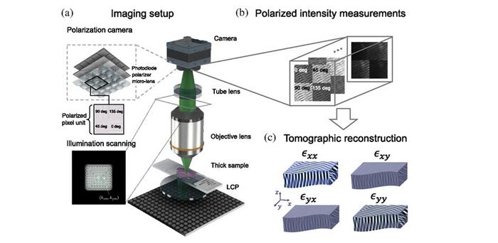
One key feature of T2oFu is its inexpensive optical setup. The system comprises an individually addressable LED matrix as the illumination source. To obtain polarization-dependent information, the system also employees a circular polarizer between the illumination and the sample, as well as a polarization-sensitive camera. To reconstruct polarization-sensitive quantitative phase tomography with this setup, the research team developed T2oFu’s reconstruction model from the ground up. Based on theories of light propagation, they derived a mathematical model that accurately describes experimental measurements.
With the experimental setup and the theoretical framework established, the team put their method to the test through a series of experiments. First, they reconstructed detailed 3D images of muscle fibers with anisotropy and phase information, obtaining a clear view of individual muscle filaments. This has important implications for diagnostic purposes. “High-contrast and high-resolution structural imaging of intrinsic signals in skeletal muscle fibers is important for the timely detection of changes in myofibrillar organization that can lead to skeletal myopathies,” explains Dr. Horstmeyer. “Currently, 3D muscle tissue is typically imaged by complex and expensive systems, such as second-harmonic generation (SHG) microscopy. Notably, our inexpensive, LED-based system showed results similar to those described in the literature on SHG imaging.”
Then, the researchers imaged a heart tissue sample with cardiac amyloidosis, a highly lethal disease that affects over 12,000 patients in the USA alone. “In current practice, biopsied heart tissue is first frozen and thinly sliced, then stained with a red-colored dye and inspected under a cross-polarized microscope,” comments Dr. Xu. “In our measurements, the structure of the anisotropy reconstruction was highly correlated with the color-stained cross-polarized image depicting features of amyloidosis. Thus, the proposed approach may potentially be useful for rapid on-site inspections in the future.”
Overall, T2oFu appears to be a powerful and practical technique that could make polarization and phase imaging more readily accessible. Further refinements will hopefully make this tool available to more scientists and doctors, lighting the way to better diagnostics and a deeper understanding of our bodies.
For details, see the original Gold Open Access article by Xu et al., “Tensorial tomographic Fourier ptychography with applications to muscle tissue imaging,” Adv. Photon. 6(2) 026004 (2024), doi 10.1117/1.AP.6.2.026004.
Journal
Advanced Photonics
DOI
10.1117/1.AP.6.2.026004
Article Title
Tensorial tomographic Fourier ptychography with applications to muscle tissue imaging
Article Publication Date
4-Mar-2024


