
In a groundbreaking study that delves into the plasticity of spatial navigation systems, researchers have uncovered new insights into how larval zebrafish encode directional information through their head direction (HD) neurons. The work challenges long-standing assumptions about the rigidity of visual landmark mapping within neural circuits responsible for navigation, revealing an experience-dependent mechanism that reshapes our understanding of sensory integration in the brain’s compass system.
Traditional models of HD neurons have posited the existence of a “ring attractor” mechanism, where a stable bump of activity within a neural network represents the animal’s orientation in space. This bump’s phase uniquely aligns with visual landmarks, learned through consistent co-activation patterns between visual neurons—each tuned to a particular part of the environment—and the HD neurons themselves. This mapping is thought to arise via Hebbian plasticity, strengthening synaptic connections when visual cues and HD neuron activity coincide repeatedly during exploration.
The researchers tested the stability and adaptability of this system by experimentally inducing a symmetric scene, introducing a twofold point symmetry to the fish’s visual environment. Instead of a single, unique landmark cue, the fish were presented with two identical visual stimuli positioned 180 degrees apart. The hypothesis was that this symmetry would disrupt the uniquely learned mapping, forcing the activity bump in HD neurons to oscillate between two diametrically opposite orientations.
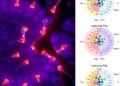
This experimental design contrasted three phases: a pre-learning epoch where a single, Sun-like visual cue was present; a learning epoch during which a second symmetrical cue was introduced; and a post-learning epoch reverting to the single-cue scenario. Through sophisticated neural recording and analytic techniques, including sinusoidal fitting to identify HD cells based on half of the pre-learning data, the researchers discerned clear shifts in response dynamics correlated with the visual experience.
Before the introduction of the symmetric scene, the HD neurons showed strong alignment between the bump phase and the visual landmark bearing, indicated by a narrowly peaked distribution centered around zero phase offset. However, during the learning phase with two suns, this alignment became variable, with the bump phase shifting unpredictably between opposite points on the ring attractor—highlighting an instability that undermines the unique mapping.
Notably, this variability persisted into the post-learning epoch even though the visual environment had returned to a single cue. The offset distribution widened significantly, suggesting that the network had incorporated the conflicting inputs into its synaptic architecture. The data indicate that the visual-to-HD neuron connectivity had effectively doubled, enabling the bump to be driven to two opposite phases by a single landmark, thus degrading the scene-bump alignment essential for reliable orientation.
Crucially, control experiments where fish were only exposed to the single landmark throughout showed no such disruption, underscoring that the effects are dependent on the animal’s visual experience and not merely time or repeated exposure. This provides strong empirical support for the idea that the landmark anchoring in the HD system is not fixed but plastic and shaped over time by sensory history.
The findings extend our knowledge of how neural circuits underlying spatial navigation balance stability and flexibility. While the ring attractor provides a robust representation of heading direction, this network remains responsive to environmental complexities, including ambiguous or symmetric visual inputs. The plasticity uncovered here may represent a fundamental biological strategy to overcome conflicting sensory cues by adjusting synaptic weights to integrate or segregate inputs as necessary.
The study suggests that the visual-to-HD synapses undergo dynamic remodeling to encode consistent spatial relational information. However, when faced with symmetrical visual information that could map onto multiple phases, the system adapts by creating duplicated synaptic pathways, which unfortunately can impair the specificity of landmark anchoring.
These results also carry important implications for understanding how animals navigate natural environments, which often contain symmetric features or repetitive patterns. The observed plasticity might underlie behavioral flexibility, enabling animals to recalibrate their internal compass when confrontations with ambiguous cues occur, yet at the cost of increased uncertainty or bistability in directional representation.
Beyond zebrafish, this research offers a conceptual framework for exploring similar plasticity mechanisms in mammalian navigation systems, including the extensive network of HD cells identified in rodents and primates. It raises the possibility that experience-dependent tuning of spatial mapping is a conserved feature across species, potentially contributing to phenomena like reorientation and spatial memory updating.
Technically, the study integrates sophisticated neural recording with computational modeling, confirming that a ring attractor network endowed with Hebbian plasticity can replicate the observed phenomena. This combined approach bridges experimental and theoretical neuroscience, deepening our mechanistic insight into how neural circuits evolve during learning and adapt to environmental challenges.
The investigators’ methodical analysis of the scene–bump offset dynamics revealed a key metric for quantifying the degree of disruption induced by scene symmetry. Tracking time spent in “out-of-phase” states—where the offset exceeds 135 degrees—served as a sensitive indicator of the breakdown in unique landmark anchoring, laying groundwork for future experimental and modeling endeavors.
In sum, this study illuminates the remarkable adaptability of compass neurons in larval zebrafish, demonstrating that visual landmark anchoring is not a fixed, static process but one shaped continuously by visual experience. Such plasticity optimizes navigation and spatial recognition amidst challenging sensory landscapes, thereby enriching our understanding of the fundamental neural computations underlying orientation and cognitive mapping.
Subject of Research: Neural plasticity in head direction neurons and visual landmark mapping in larval zebrafish
Article Title: Plastic landmark anchoring in zebrafish compass neurons
Article References:
Tanaka, R., Portugues, R. Plastic landmark anchoring in zebrafish compass neurons. Nature (2026). https://doi.org/10.1038/s41586-025-09888-x
Image Credits: AI Generated
DOI: https://doi.org/10.1038/s41586-025-09888-x
Tags: directional information encoding in fishexperience-dependent neural mechanismsgroundbreaking research on zebrafishhead direction neurons in zebrafishHebbian plasticity in navigationneural circuits in spatial orientationplasticity of spatial navigationsensory integration in navigationstability of neural networkssymmetry in visual stimulivisual landmark mapping in fishzebrafish navigation mechanisms

