Credit: by La Cavera, S., Pérez-Cota, F., Smith, R.J. et al.
Ultrasound is an indispensable tool for the life sciences and various industrial applications due to its non-destructive, high contrast, and high resolution qualities. A persistent challenge over the years has been how to increase the resolution of an acoustic endoscope without drastically increasing the footprint of the probe, or risking the robustness of the ultrasonic transducer. In recent years, a host of all-optical ultrasonic imaging techniques have emerged – which generally utilise pulsed lasers and optical cavities to excite and detect ultrasound waves – without sacrificing device footprint, sensitivity, or the integrity of the transducer. Thus far these powerful techniques have achieved imaging resolutions on microscopic-mesoscopic length scales, however there is great interest in creating an ultrasonic fibre-probe which can probe disease on the nanoscopic-microscopic length scales inhabited by biological cells and tissue.
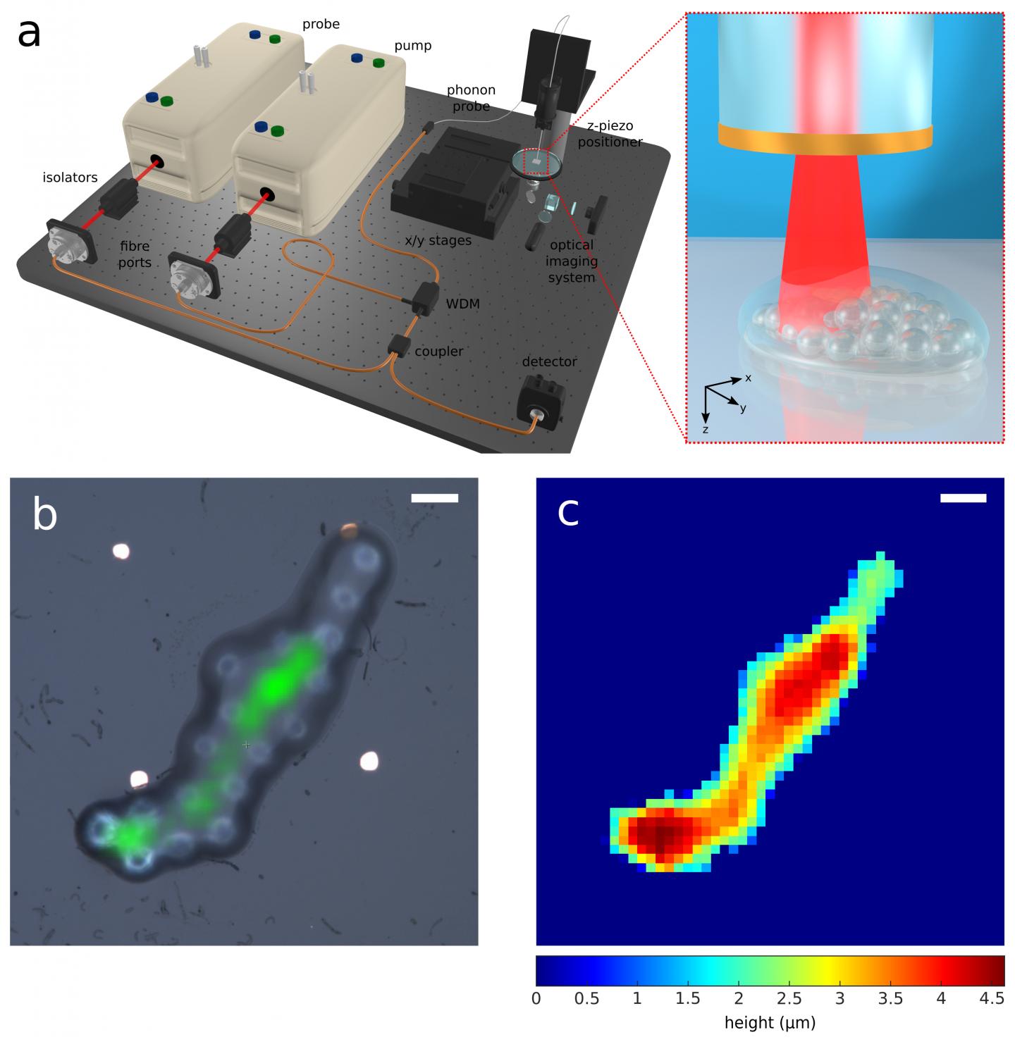
In a new paper published in Light: Science & Application, a team of scientists, led by Professor Matt Clark at the University of Nottingham, UK, has developed the first optical fibre ultrasonic imaging device which operates in the GHz range of the acoustic spectrum. At these frequencies the wavelength of sound becomes comparable to ultraviolet optical wavelengths and therefore provides an opportunity for high resolution imaging. Their phonon probe device makes use of a pump-probe technique called time-resolved Brillouin scattering, which pumps GHz frequency ultrasound from the tip of a 125 μm diameter optical fibre into a specimen, and uses a pulsed laser to “watch” one of the ultrasound waves (with a frequency of approximately 5 GHz) as it travels through the specimen. This time-of-flight acoustic signature simultaneously encodes two types of information about the specimen: its local mechanical properties and spatial profile. By scanning the device, these properties can be resolved in 3D with optical lateral resolution, and with axial resolution dictated by the sub-micrometre acoustic wavelengths.
Prof. Clark’s team applied this new technology to the parallel elastography-profilometry of objects as small as 10 x 2 μm (radius and height). The device was capable of 2.5 μm lateral resolution and could measure object height with 45 nm precision, which is over an order of magnitude smaller than the system’s optical wavelength. The team also demonstrated that the technology is fully compatible with optical fibre imaging bundles – containing tens of thousands of imaging pixels – which shows the scalability of the technique and its compatibility with standard endoscopy equipment.
According to the team, the development and application towards biological metrology and healthcare is most exciting. “The phonon probe is poised to supplement state-of-the-art bench-top profilometry equipment such as atomic force microscopy (AFM), stylus profilometry, and optical profilometry. It offers a combination of non-contact operation, label-free contrast, and high resolution, which is unique compared with the state-of-the-art. However, we believe that its ability to measure sub-surface mechanical properties, its bio-compatibility, and its endoscopic-potential are what set it apart. These features set the technology up for future in vivo measurements towards the ultimate goal of minimally invasive point-of-care diagnostics. The building blocks of disease can be traced down to the sub-cellular level, and are closely intertwined with mechanical properties. Having an endoscopic device that can access this regime will accelerate the development of elasticity-based diagnostics.”
###
Media Contact
Salvatore La Cavera III
[email protected]
Related Journal Article
http://dx.


