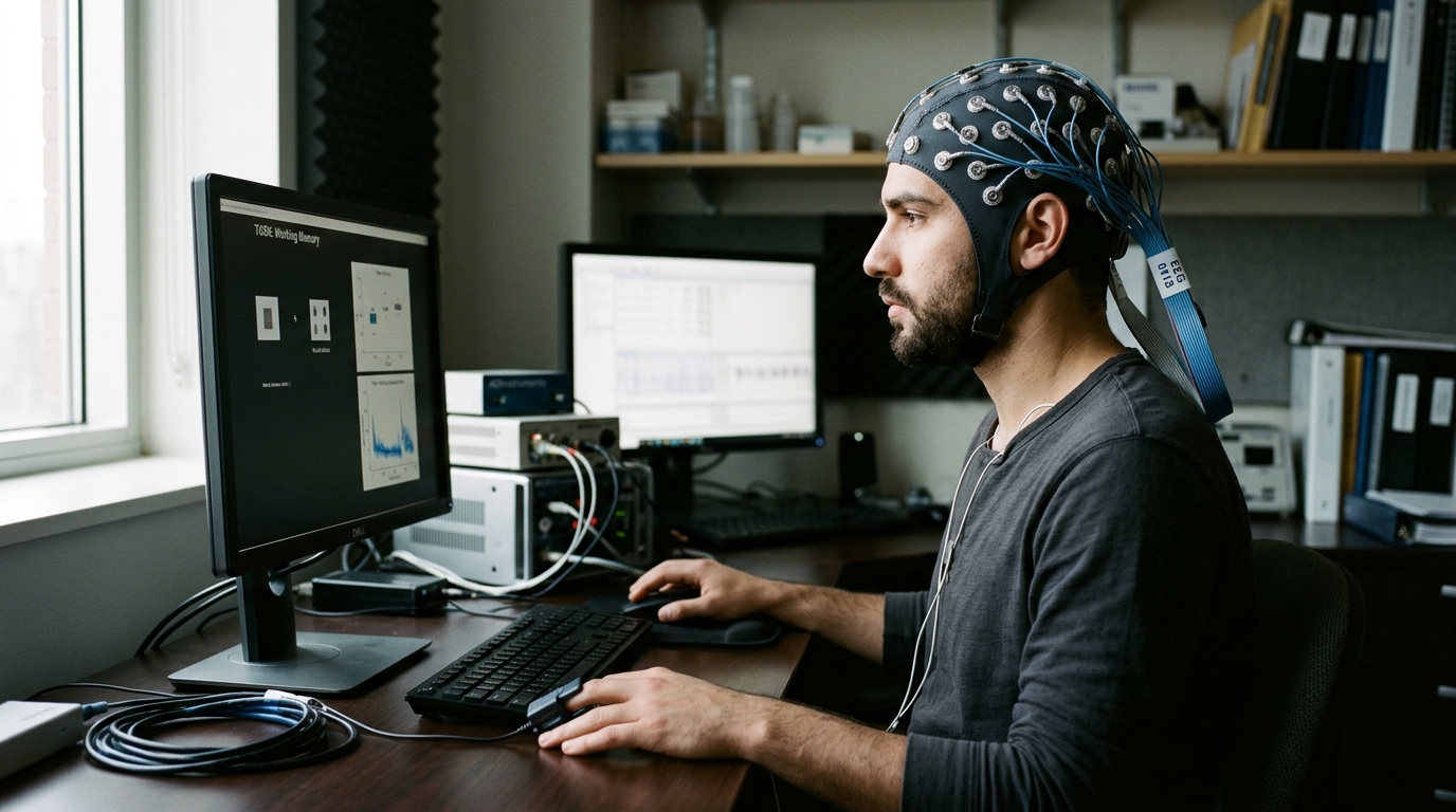
A research team led by Matteo Saibene and Sadasivan Puthusserypady from the Technical University of Denmark, collaborating with clinical specialists from the University of Copenhagen and Zealand University Hospital, has investigated directional brain-heart interactions during mental arithmetic using permutation conditional mutual information. Brain-heart interaction reflects the dynamic coupling between cortical activity and autonomic cardiovascular regulation, functioning as an observable expression of the brain-heart axis. Understanding how neural activity and autonomic regulation communicate bidirectionally holds important implications for neurology, affective neuroscience, sleep research, and clinical risk evaluation. To capture directional dynamics under cognitive workload, the authors evaluated directed dependencies between electroencephalography and electrocardiography signals. Applying their computational framework to simultaneous recordings obtained during resting baseline and structured subtraction tasks, the researchers demonstrated that cognitive workload significantly increases the temporal complexity of directional coupling across cortical regions and autonomic frequency bands.
The physiological architecture governing brain-heart communication comprises an intricate network of ascending and descending anatomical pathways linking the central nervous system with peripheral autonomic effectors. Autonomic cardiac control is mediated by a multilevel system involving intrathoracic neuronal clusters, extrathoracic ganglia, and tiers of the central autonomic network spanning the spinal cord, brainstem, and basal ganglia. At supranuclear levels, cerebral influence over cardiovascular function operates primarily through the central autonomic network, incorporating the insular cortex, amygdala, medial prefrontal cortex, lateral hypothalamus, and medullary centers. Within the medulla, neuronal circuits in the nucleus tractus solitarius and ventrolateral medulla regulate blood pressure, respiration, and heart rate. While reciprocal brain-heart variations occur across sleep, emotional elicitation, and meditation, demanding cognitive tasks provide an effective experimental model for examining how mental effort reorganizes large-scale cortical networks and autonomic cardiovascular dynamics.
Investigating brain-heart interactions through electroencephalography and electrocardiography presents substantial methodological challenges because physiological time series are inherently non-stationary, noisy, and nonlinearly coupled. Existing analytical frameworks—including heartbeat evoked potentials, linear Granger causality, time delay stability, convergent cross mapping, and synthetic generation models—often struggle to capture nonlinear, time-varying interactions across simultaneous multivariate channels. To overcome these constraints, the authors examined permutation conditional mutual information, combining conditional mutual information with permutation entropy. This formulation evaluates directional information transfer between two observables by transforming continuous time series into discrete ordinal patterns based on the ranking of neighboring values within embedded segments. Because ordinal mapping depends on relative ordering rather than absolute amplitudes, the method provides increased robustness to low-frequency recording drift and amplitude fluctuations, while effectively capturing nonlinear directional dependencies and temporal coupling asymmetries.
Before analyzing human experimental recordings, the investigators evaluated the permutation conditional mutual information directionality index on synthetic coupled time series using linear autoregressive processes and nonlinear van der Pol oscillators. Across coupled autoregressive simulations with coupling strengths ranging from zero to one over one hundred repetitions, the directionality index reliably tracked imposed coupling strengths, stabilizing once coupling exceeded 0.5. Tests under additive noise with signal-to-noise ratios from negative ten to twenty decibels showed that strong noise contamination compromised directional detection, whereas higher signal-to-noise ratios produced stable coupling estimates. Complementary simulations with coupled van der Pol oscillators using a damping parameter of one confirmed that systematically varying asymmetrical coupling coefficients shifted the directionality index toward the dominant driving oscillator, verifying the method's ability to recover directional coupling between nonlinear dynamical systems.
The clinical analysis utilized the publicly available EEG During Mental Arithmetic Tasks dataset from PhysioNet, containing simultaneous nineteen-electrode electroencephalography and single-lead electrocardiography recorded at five hundred hertz across thirty-six age-matched participants with a mean age of eighteen years. The cohort comprised twenty-seven females and nine males with normal vision and no history of neurological impairment, psychiatric illness, or psychoactive medication use. Based on subtraction accuracy and operations completed during a four-minute task, the original dataset classified twenty-four participants as good counters, averaging twenty-one operations, and twelve participants as bad counters, averaging seven operations. Preprocessing applied 0.5 to 45 hertz band-pass filtering and a 50 hertz notch filter to cortical channels before independent component analysis was executed to attenuate ocular, muscular, and cardiac pulsation artifacts. Continuous interbeat interval series extracted from electrocardiography were resampled at one hertz for spectral processing.
Feature extraction was performed independently for each signal modality to generate matched time series with uniform one-second temporal sampling. Cortical power spectral density was estimated across nineteen scalp electrodes using short-time Fourier transforms with one-thousand-sample Hanning windows and fifty percent overlap, yielding power values integrated within delta, theta, alpha, beta, and gamma bands. Heart rate variability features were derived from resampled interbeat intervals using smoothed pseudo Wigner-Ville distributions with 256 frequency bins, integrating power across low-frequency bands from 0.04 to 0.15 hertz and high-frequency bands from 0.15 to 0.40 hertz. The high-frequency component primarily reflects vagal parasympathetic activity, whereas low-frequency power represents mixed autonomic and baroreflex dynamics. Heart rate variability features were corrected for heart rate before computing time-varying permutation metrics, which were calculated in sliding ten-second windows with fifty percent overlap using an embedding dimension of three and unit time delay.
To quantify the temporal regularity and dynamical complexity of the directionality index series, the researchers calculated fuzzy entropy across all electrode-frequency pairings. Fuzzy entropy evaluates time series regularity using exponential membership functions controlled by vector dimension, boundary gradient, and tolerance width parameters, where higher entropy values indicate greater dynamical complexity. Parameter optimization on a random data subset established specific values for the fuzzy entropy parameters, which were held constant across all subjects. Statistical differences between baseline and arithmetic tasks were assessed using paired two-tailed cluster-based nonparametric permutation tests with five thousand permutations. Clusters were formed using an uncorrected threshold of p equals 0.05, and multiple comparisons across the ten electroencephalographic and heart rate variability coupling pairs were controlled using Bonferroni correction, setting the significance threshold at p less than 0.005.
The empirical findings revealed that directional brain-heart coupling entropy increased significantly during mental arithmetic compared to resting baseline, demonstrating substantial effect sizes with Cohen’s d values ranging from 0.56 to 1.82 across significant electrode clusters. This entropy increase indicates elevated temporal irregularity and complexity in directional brain-heart communication during cognitive effort. Topographical distributions showed significant effects extending broadly across anterior, temporal, parietal, and occipital scalp regions rather than remaining confined to localized cortical areas. Couplings involving high-frequency heart rate variability exhibited significant entropy shifts across delta, theta, and beta bands, with prominent temporal electrode involvement reflecting task-related changes in vagal cardiovascular modulation. Simultaneously, low-frequency cardiac couplings showed significant differences across anterior and posterior regions, particularly within the alpha band. These widespread patterns suggest that mental workload recruits distributed cortical networks, altering both vagally mediated loops and mixed baroreflex cardiovascular regulation.
Beyond evaluating temporal complexity through fuzzy entropy, the researchers examined the signed group mean of the directionality index to determine whether cognitive workload reversed the dominant direction of information transfer. In their formulation, positive values denote predominant brain-to-heart signaling, whereas negative values indicate predominant heart-to-brain driving. Across nine of the ten coupling pairs, the sign of the group mean directionality index remained unchanged between baseline and mental arithmetic conditions, indicating preserved dominant information transfer directions. The only pairing exhibiting a directional reversal was low-frequency heart rate variability coupled with cortical gamma activity, shifting from predominant heart-to-brain driving during baseline to brain-to-heart directionality during arithmetic. Crucially, the authors emphasized that directionality signs reflect predominant predictive direction rather than physiological causality, and that elevated entropy captures temporal irregularity in the directionality series rather than an increase in overall coupling strength.
When participants were stratified into good and bad counters based on calculation performance, between-group comparisons revealed no statistically significant differences in coupling entropy during the mental arithmetic task. Normalized entropy values remained largely comparable across both performance groups, although visual inspection identified a localized positive cluster over posterior scalp regions for low-frequency cardiac coupling to cortical alpha rhythms. However, this effect did not survive cluster-based multiple comparison correction, demonstrating that directional coupling complexity reflects general cognitive workload rather than arithmetic success. Substantial inter-individual variability was observed across all coupling pairs, with descriptive subject-level absolute entropy differences between baseline and task ranging from 0.6989 to 0.8524 across both performance categories. This variability suggests that while cognitive effort broadly elevates brain-heart interaction complexity across subjects, individual physiological traits, attentional allocation, and autonomic regulation generate diverse individual response profiles.
The authors outlined several methodological limitations that contextualize their results and delineate directions for future research. The study cohort was relatively small, consisting of thirty-six young participants with an average age of eighteen years and a significant female predominance, limiting immediate generalizability to broader demographic or clinical populations. Furthermore, the protocol utilized only a mental subtraction task, which may not capture autonomic responses elicited by other cognitive paradigms. Scalp electroencephalography without source reconstruction restricted interpretations to sensor-level patterns rather than deep structures of the central autonomic network, and residual cardiac field artifacts could not be fully excluded. Additionally, ten-second estimation windows reflected a trade-off between temporal resolution and sample reliability, and respiration was not monitored, precluding exclusive attribution of high-frequency heart rate variability to vagal tone. Nevertheless, permutation conditional mutual information offers a promising nonlinear framework for investigating brain-body interactions across network physiology.
Subject of Research: Directional brain-heart interaction during cognitive workload
Article Title: Directional brain-heart interaction during cognitive workload: a permutation conditional mutual information approach
Article References: Saibene, M., Ballegaard, M., Andersen, T. S., Gu, Y., Bardram, J. E., & Puthusserypady, S. (2026). Directional brain-heart interaction during cognitive workload: a permutation conditional mutual information approach. Frontiers in Network Physiology, 6, Article 1930070. https://doi.org/10.3389/fnetp.2026.1930070
Image Credits: AI Generated
DOI: 10.3389/fnetp.2026.1930070
Keywords: brain-heart interaction, electroencephalography, heart rate variability, permutation conditional mutual information, fuzzy entropy, cognitive workload, central autonomic network, network physiology, Directional, brain-heart, interaction, during
News Source: Cassandra Pierce. (November 7, 2026). Permutation Conditional Mutual Information Uncovers Directional Brain-Heart Coupling Under Cognitive Workload. Scienmag.