
In a groundbreaking study published in Cell Death Discovery, researchers have unveiled critical insights into the cellular mechanisms that exacerbate diabetic kidney disease (DKD), focusing on the pivotal role of p16-positive senescent cells. This research dissects how these senescent cells trigger a cascade of metabolic disturbances, specifically through the dysregulation of glycolysis and mitochondrial function, shedding light on previously obscure pathways that underpin the progression of DKD. The implications resonate profoundly for therapeutic strategies targeting cellular senescence and metabolic reprogramming in chronic kidney diseases.
Senescence, a state of irreversible cell cycle arrest, is increasingly recognized not only as a hallmark of aging but also as a driver of various chronic pathologies, including diabetic complications. P16^INK4a, a cyclin-dependent kinase inhibitor, is a well-established biomarker marking senescent cells, which accumulate in tissues under metabolic stress such as those observed in diabetes. The study profoundly connects the dots between the accumulation of these p16-positive cells and the perturbations in energy metabolism that fuel DKD progression.
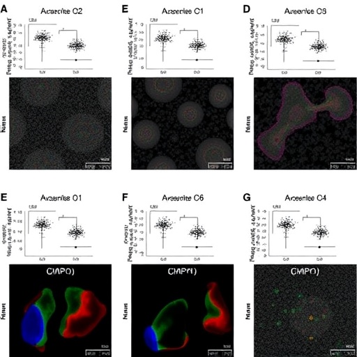
The team led by Lu, X., and colleagues utilized advanced biochemical and molecular biology techniques to investigate the bioenergetic profiles of renal cells harboring p16-induced senescence. Their experiments revealed that senescent cells exhibit an impaired glycolytic pathway accompanied by mitochondrial dysfunction, which collectively compromise cellular energy homeostasis. This metabolic imbalance not only undermines cellular viability but also ignites pro-fibrotic and pro-inflammatory signaling pathways, potentially accelerating kidney damage in diabetic milieus.
.adsslot_Fp7zfV5NxH{width:728px !important;height:90px !important;}
@media(max-width:1199px){ .adsslot_Fp7zfV5NxH{width:468px !important;height:60px !important;}
}
@media(max-width:767px){ .adsslot_Fp7zfV5NxH{width:320px !important;height:50px !important;}
}
ADVERTISEMENT
Intriguingly, the researchers noted an aberrant shift in glycolytic flux, characterized by diminished conversion of glucose to pyruvate and a concomitant decrease in ATP generation. This attenuation of glycolysis was mirrored by mitochondrial respiratory defects, including altered membrane potential and reduced oxidative phosphorylation capacity. Such mitochondrial anomalies further exacerbate oxidative stress and promote the secretion of senescence-associated secretory phenotype (SASP) factors, which propagate tissue inflammation and fibrosis, hallmark features of DKD.
The meticulous examination of senescence markers alongside metabolic enzyme expression profiles underscored a tightly interwoven relationship between cell cycle arrest and energy metabolism. The interplay suggests that p16 expression not only demarcates senescence but actively orchestrates metabolic reprogramming, placing mitochondrial and glycolytic dysfunctions at the epicenter of DKD pathogenesis.
One particularly compelling element of this research lies in its potential clinical translatability. By delineating the metabolic fingerprint of p16-positive senescent cells, therapeutic avenues targeting these dysfunctional pathways come into sharper focus. Modulating glycolysis or restoring mitochondrial integrity could mitigate the deleterious effects of senescent cells, offering new hope for patients grappling with the relentless advance of diabetic nephropathy.
Moreover, the findings advocate for a paradigm shift in how diabetic kidney disease is approached—from primarily glucose-centric strategies to interventions that address the intricate cellular senescence and metabolic disruptions. This expanded conceptual framework paves the way for combination therapies that could simultaneously suppress senescence-associated signaling and restore metabolic balance.
The study’s use of state-of-the-art assays to quantify changes in glycolytic intermediates and mitochondrial respiration highlights the crucial role of integrated bioenergetic profiling in understanding disease mechanisms. These technological advancements enabled the identification of precise metabolic nodes altered in senescent cells, providing a granular view that was previously unattainable.
Crucial to this endeavor was the characterization of the senescence-associated secretory phenotype, which elucidates how senescent cells influence the renal microenvironment. The release of inflammatory cytokines, chemokines, and growth factors from p16-positive cells fosters a vicious cycle of tissue remodeling and dysfunction, which was detailed elegantly in this study.
The authors also shed light on potential molecular targets within these metabolic pathways. Enzymes regulating key glycolytic steps and mitochondrial complexes represent strategic nodes that could be pharmacologically manipulated to reverse or alleviate the senescent phenotype and its pathological consequences.
Importantly, this research integrates findings from cellular models with analyses of kidney tissues from diabetic patients, reinforcing the translational relevance and underscoring the universality of the observed metabolic alterations. Such congruence between model systems and human pathology bolsters the confidence in targeting these pathways clinically.
The investigation’s scope extended beyond metabolic characterization, delving into the signaling cascades initiated by senescence-driven metabolic dysfunction. These pathways feed into fibrotic processes and immune system dysregulation, both pivotal in the progression of diabetic nephropathy. Understanding these interactions opens new vistas for multifaceted therapeutic interventions.
By juxtaposing metabolic dysregulation with the phenotypic manifestations of DKD, the research collectively paints a comprehensive picture of disease progression. The nuanced elucidation of how energy metabolism intertwines with cellular aging mechanisms provides a rich framework to decode the complexity of diabetic kidney damage.
Looking forward, the authors propose that future research should explore senolytic or senostatic drugs that specifically target p16-positive cells, in combination with agents that restore metabolic competence. Such a two-pronged approach could effectively halt or even reverse diabetic kidney disease progression.
In conclusion, this study marks a significant advance in our understanding of the cellular and metabolic underpinnings of diabetic kidney disease. It spotlights p16-positive senescent cells as key pathological players whose metabolic disturbances catalyze kidney damage. This work not only enriches the scientific narrative surrounding DKD but also lays the groundwork for innovative therapeutic strategies that could transform patient outcomes in this pervasive and debilitating disease.
Subject of Research: The role of p16-positive senescent cells in promoting diabetic kidney disease through the dysregulation of glycolysis and mitochondrial metabolism.
Article Title: P16-positive senescent cells promote DKD by the dysregulation of glycolysis and mitochondrial metabolism.
Article References:
Lu, X., Wu, J., Agborbesong, E. et al. P16-positive senescent cells promote DKD by the dysregulation of glycolysis and mitochondrial metabolism.
Cell Death Discov. 11, 355 (2025). https://doi.org/10.1038/s41420-025-02650-2
Image Credits: AI Generated
DOI: https://doi.org/10.1038/s41420-025-02650-2
Tags: advanced biochemical techniques in researchbiomarkers of cellular senescencecellular senescence in chronic kidney diseasesdiabetic kidney disease metabolic dysfunctionenergy metabolism in kidney diseaseglycolysis dysregulation in DKDimplications of senescence in diabetesirreversible cell cycle arrest in agingmetabolic reprogramming in renal healthmitochondrial function and senescencep16-positive senescent cellstherapeutic strategies for diabetic complications

