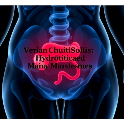
The medical community continues to grapple with the perplexing and rarely encountered condition known as ovarian hydatidosis, a manifestation of echinococcosis that poses significant diagnostic and therapeutic challenges. In a groundbreaking study recently published in Acta Parasitologica, Iranian researchers Hezarjaribi, Soleymani, Ghahghaei-Nezamabadi, and colleagues have dissected the complexities surrounding this parasitic infection, offering new insights into its presentation, diagnosis, and management. This condition, stemming from echinococcus tapeworm larvae, is notoriously difficult to identify, often masquerading as other ovarian pathologies, which can delay proper treatment and complicate patient outcomes.
Echinococcosis primarily affects the liver and lungs; however, its rare involvement of ovarian tissue represents an under-recognized clinical entity. The difficulty lies in its mimicry of more common ovarian cysts or tumors on imaging and clinical examination. The researchers emphasize that ovarian hydatidosis should enter differential diagnoses, particularly in endemic regions like Iran, with its elevated rates of echinococcal infections. The study’s detailed case report exemplifies the typical clinical course, including the radiological and laboratory findings that initially misled the diagnostic process.
The report underscores the role of advanced imaging modalities in suspecting hydatid cysts. Ultrasound and computed tomography scans may reveal cystic lesions, yet their features often overlap with other cystic ovarian masses. Particular imaging characteristics—multiloculated cysts with daughter cysts—suggest hydatid disease but are far from pathognomonic. Magnetic resonance imaging (MRI), with its superior soft tissue resolution, may aid subtle differentiation but remains insufficient as a standalone tool. Hence, radiologic interpretation demands expertise and the inclusion of epidemiological context.
Serological testing for echinococcal antibodies introduces another layer of diagnostic complexity. Though serology can confirm exposure, false negatives and cross-reactivities are common. The authors note that the sensitivity and specificity of these tests vary widely, influenced by cyst location, stage, and host immune response. Therefore, serological assays must be interpreted cautiously and in conjunction with imaging and clinical data. The necessity for improved, more reliable diagnostic biomarkers is evident from these findings.
Surgical intervention remains the cornerstone of treatment for ovarian hydatidosis, yet it is fraught with inherent risks. The distinct possibility of cyst rupture during manipulation threatens to disseminate parasitic material intraoperatively, causing severe anaphylactic reactions and secondary hydatidosis. Surgeons must employ meticulous techniques, often under the cover of antiparasitic chemotherapy, to mitigate these dangers. The authors detail their case’s operative approach, highlighting the delicate balance between complete cyst excision and preservation of ovarian tissue to maintain fertility.
Adjunctive therapy with benzimidazoles such as albendazole is critical in the multidisciplinary management of ovarian hydatidosis. Preoperative administration aims to sterilize cyst contents, reducing the risk of anaphylaxis and recurrence. Postoperative therapy addresses residual disease and prevents new cyst formation. However, drug regimens are frequently prolonged and associated with side effects, requiring vigilant monitoring of liver function and hematologic parameters. The article elaborates on optimizing antiparasitic protocols tailored to individual patient profiles, underscoring the need for personalized medicine.
The authors’ exhaustive literature review reveals a paucity of robust data on the epidemiology and long-term outcomes of ovarian hydatidosis, stressing a gap in global parasitic disease knowledge. Most documented cases stem from endemic regions in the Middle East and Central Asia, where healthcare infrastructure may be limited. This scarcity of comprehensive research restricts clinicians’ ability to develop standardized diagnostic algorithms and therapeutic guidelines, perpetuating variability in patient care and outcomes. Collaborative international efforts are imperative to bridge these gaps.
The study further highlights the psychosocial burden imposed by this obscure disease. Patients often endure prolonged diagnostic odysseys compounded by fears of malignancy due to the cystic presentation of ovarian lesions. The stigma associated with parasitic infections and uncertainty about fertility outcomes exacerbate distress. Incorporating psychological support within the clinical pathway is a noteworthy recommendation from the authors, reflecting a holistic approach to patient wellbeing seldom emphasized in parasitic disease management.
Epidemiologists and public health officials should also take note of the zoonotic transmission pathways implicating domestic and wild canids as definitive hosts of echinococcus species. Improved control measures targeting these reservoirs, including deworming programs and sanitation improvements, could reduce human infection incidence. Health education campaigns tailored to at-risk populations would further enhance early detection and intervention. This study reinforces the interconnectedness of veterinary, human, and environmental health within the One Health paradigm.
On a molecular level, the research team calls for intensified investigation into the genetic and immunological interactions underpinning ovarian hydatidosis. Understanding host immune evasion mechanisms employed by echinococcus larvae can unearth novel therapeutic targets. Advances in proteomics and immunogenetics might facilitate the development of vaccines or targeted biologics, revolutionizing the prophylaxis and treatment of this neglected condition.
Moreover, artificial intelligence and machine learning hold promise in refining diagnostic accuracy. Integrating clinical, imaging, serological, and molecular data could yield predictive algorithms capable of stratifying hydatid cyst risks and guiding management decisions. The authors speculate that future integration of such technologies could transform care pathways, minimizing unnecessary interventions and expediting diagnosis.
The case report exemplifies the intricate interplay between parasitology, gynecology, surgery, and infectious diseases. Coordinated multidisciplinary teams, including radiologists, pathologists, and pharmacists, are vital to navigating the diagnostic labyrinth and delivering effective therapy. This collaborative approach is essential in endemic areas to combat the morbidity associated with ovarian hydatidosis and improve survival rates.
In conclusion, Hezarjaribi and colleagues’ work illuminates the enigmatic presentation of ovarian hydatidosis, pushing the medical community to consider this entity more vigorously in differential diagnoses and to refine management strategies. Their meticulous case documentation and comprehensive literature synthesis provide a valuable framework that can inspire future research, enhance clinical practice, and ultimately improve patient outcomes in a neglected yet impactful parasitic disease realm.
As the global burden of parasitic diseases continues to evolve, studies like this underscore the importance of vigilance, innovation, and collaboration in tackling rare but significant infections. Ovarian hydatidosis, once shrouded in mystery and misdiagnoses, now emerges as a beacon calling for enhanced awareness, research investment, and healthcare synergy worldwide.
Subject of Research: Ovarian Hydatidosis – Diagnosis and Management Challenges
Article Title: Challenges in Diagnosing and Managing Ovarian Hydatidosis: A Case Report and Literature Review from Iran
Article References:
Hezarjaribi, H.Z., Soleymani, E., Ghahghaei-Nezamabadi, A. et al. Challenges in Diagnosing and Managing Ovarian Hydatidosis: A Case Report and Literature Review from Iran. Acta Parasit. 70, 237 (2025). https://doi.org/10.1007/s11686-025-01177-x
Image Credits: AI Generated
DOI: https://doi.org/10.1007/s11686-025-01177-x
Tags: advanced imaging in parasitologycase studies in rare diseaseschallenges in diagnosing ovarian pathologiesclinical presentation of hydatid cystscomputed tomography in parasitic infectionsdifferential diagnosis of ovarian massesechinococcosis management challengesendemic regions and echinococcal infectionsimaging techniques for ovarian cystsovarian hydatidosis diagnosisparasitic infections in womenultrasound in ovarian diagnostics

