
Credit: Salk Institute
LA JOLLA–(April 24, 2018) Glioblastoma multiforme (GBM) is an incredibly deadly brain cancer and presents a serious black box challenge. It's virtually impossible to observe how these tumors operate in their natural environment and animal models don't always provide good answers.
But now, Salk Institute researchers have taken an important step towards meeting that challenge. By editing two genes in just a few cells in human cerebral organoids, scientists in the Verma lab generated aggressive GBM tumors. This new model could be used to study tumor progression, investigate new drugs or even personalize treatments for patients. The study was published in the journal Cell Reports on April 24, 2018.
One of the problems plaguing clinical trials is, quite often, drugs that work in animals do not work in people. Researchers have tried to overcome this by using xenografts, in which patient tumor tissue is implanted in animal models, but this approach has its own issues. Sometimes, there isn't enough human tumor tissue to study and, over time, the tumors adapt to their new home.
"As tumors grow in mice, the environment changes the tumor's features," says Junko Ogawa, a Salk senior research associate and first author on the paper. "We don't know if it's similar to the patient's original cancer."
The solution could be human cerebral organoids, which contain neurons and other brain cells. The Salk lab has been using stem cells to generate these small (around 4 mm) 3D structures in a dish for some time and wanted to investigate how they could be applied to study GBM.
They used the CRISPR-Cas9 tool to edit two genes closely associated with cancer, HRas and p53, in a few cells in an organoid. HRas is a cancer oncogene that drives rampant cell growth, while p53 is a tumor suppressor. In other words, they took their foot off the brake and stomped on the gas.
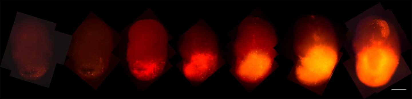
These organoids turned into tumor-like structures in the dish–they grew aggressively and had several biomarkers associated with GBM. Eventually, they took over the organoids, supplanting the original cells with tumor tissue. In addition, they could be serially transplanted into animal models, where they were also quite aggressive.
This approach offers a number of advantages. Editing p53 and HRas in just a few cells better replicates how GBMs actually develop in people–they don't start as thousands of cells at once (like a xenograft) but rather as one or two aberrant cells.
The team added a fluorescent red marker, called tdTomato, to the oncogenic HRas. As those cells took over the organoids, the researchers could track their progression. In addition, when the organoid tumors were transplanted into the brains of mice, they grew rapidly and resembled tumors taken from patients, offering easier access to samples.
"You can phenocopy the properties of the tumors in a mouse," says Ogawa, "and now we can give them drugs to see if they are effective. We can also test the tumor's ability to invade normal brain tissue."
These organoids could also host human tumor samples and some GBM cell lines. This model could be used to personalize care. Researchers and clinicians could transplant the cancer cells from patients to make organoid models. As a result, they could study how a tumor responds to treatment in cells that match the patient's genome. While the organoids lack endothelial cells and an immune system (which would give them more complexity and help them better replicate actual brain tissue), this model could be quite useful in studying a variety of brain metastatic cancers, not just GBM.
###
This work was funded by the National Institutes of Health (R01CA095613, P30 CA014195-38, P30 014195, P30 014195 and P30 014195), the H.N. and Frances C. Berger Foundation, the Leona M. and Harry B. Helmsley Charitable Trust (grant 2017-PG-MED001), the Glenn Center for Aging Research and the Chapman Foundation.
About the Salk Institute for Biological Studies:
Every cure has a starting point. The Salk Institute embodies Jonas Salk's mission to dare to make dreams into reality. Its internationally renowned and award-winning scientists explore the very foundations of life, seeking new understandings in neuroscience, genetics, immunology, plant biology and more. The Institute is an independent nonprofit organization and architectural landmark: small by choice, intimate by nature and fearless in the face of any challenge. Be it cancer or Alzheimer's, aging or diabetes, Salk is where cures begin. Learn more at: salk.edu.
Media Contact
Salk Communications
[email protected]
858-453-4100
@salkinstitute
Original Source
https://www.salk.edu/news-release/organoids-reveal-how-a-deadly-brain-cancer-grows/
