In a groundbreaking advancement that promises to redefine vascular diagnostics, researchers have introduced a pioneering method employing optoacoustic mesoscopy to address endothelial dysfunction at the level of a single capillary. This breakthrough, detailed in a recent publication in Light: Science & Applications, reveals a non-invasive, highly precise imaging modality that can resolve the minute details of vascular impairments, ushering in a new era of personalized cardiovascular medicine.
Endothelial dysfunction is widely recognized as a precursor to a myriad of cardiovascular diseases, including atherosclerosis, hypertension, and thrombosis. Traditionally, its diagnosis involves systemic assessments or indirect markers that lack the spatial resolution to isolate dysfunction within individual microvessels. The inability to examine single capillaries in situ has led to a gap in understanding the localized pathophysiology and delayed therapeutic interventions.
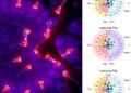
The novel approach leverages optoacoustic mesoscopy, a hybrid imaging technology that exploits the photoacoustic effect, where pulsed laser light pulses induce ultrasound waves in tissues. This technique synergistically combines the contrast advantages of optical imaging with the spatial resolution of ultrasound, enabling visualization of vascular structures as small as single capillaries at unprecedented clarity. By tuning the optical excitation wavelengths, the system can distinctly capture the absorption characteristics of hemoglobin, allowing direct visualization of blood flow and oxygenation dynamics.
The research team led by He et al. meticulously designed a single-capillary imaging system that harnesses this technology to not only detect but also quantify endothelial dysfunction. This capability stems from the innovative scanning mechanisms and signal processing algorithms that enable the differentiation between healthy and dysfunctional endothelium based on changes in capillary morphology and hemodynamic parameters. The system’s sensitivity facilitates real-time monitoring, important for understanding the progression of vascular pathologies and evaluating therapeutic responses.
Crucially, the study demonstrates that optoacoustic mesoscopy can be used to resolve endothelial dysfunction without the need for invasive angiography or contrast dyes, which often pose risks to patients and are not suitable for repeated measures. This non-destructive approach preserves the native physiological environment, allowing longitudinal studies that track capillary health dynamically, an essential factor in chronic disease management and drug efficacy tests.
The implementation of this technology required overcoming substantial technical challenges, including optimizing laser pulse energy to ensure tissue safety while maintaining signal strength, enhancing detector sensitivity, and developing sophisticated computational models to reconstruct high-resolution, three-dimensional vascular images. These innovations collectively result in a system that achieves a striking balance between spatial resolution, penetration depth, and functional imaging capability.
The clinical implications of resolving endothelial dysfunction at such a granular level are profound. Early detection of microvascular impairments can facilitate preemptive therapeutic strategies, potentially mitigating the cascade of events leading to overt cardiovascular disease. Furthermore, this imaging modality could revolutionize the screening of diabetic retinopathy, peripheral artery disease, and other conditions where microvascular integrity is compromised.
Beyond diagnostics, optoacoustic mesoscopy offers a powerful investigative tool for fundamental vascular biology. Its capacity to visualize capillary networks and endothelial responses under various physiological and pathological conditions could illuminate mechanisms of vascular remodeling, angiogenesis, and inflammation. This could catalyze breakthroughs in understanding diseases characterized by microcirculatory dysfunction, ranging from cancer to neurodegenerative disorders.
The research also highlights the translational potential of optoacoustic mesoscopy into personalized medicine. By providing a detailed vascular map for individual patients, therapies can be tailored and adjusted based on real-time feedback, increasing efficacy and reducing adverse effects. This precision approach aligns with ongoing shifts toward integrating advanced imaging with genomics and biomarker analyses.
Importantly, this study stands as a testament to multidisciplinary collaboration, synthesizing expertise in photonics, engineering, computational modeling, and vascular biology. The resulting innovation exemplifies how convergent technologies can tackle entrenched biomedical challenges, yielding tools that were previously inconceivable.
While the reported system currently excels in controlled laboratory settings, ongoing efforts focus on enhancing portability and user-friendliness to facilitate clinical adoption. Integrating this system into clinical workflows could dramatically change how vascular health is monitored, offering a powerful adjunct or alternative to existing diagnostic modalities.
The scalability of optoacoustic mesoscopy also offers promising avenues for future research and applications. Enhancements in laser sources and detector arrays could expand the field of view, enabling simultaneous imaging of multiplexed vascular networks or the coupling with functional assays to assess endothelial cell signaling in real-time.
Moreover, the underlying technology’s flexibility allows adaptation to other biological tissues and disease models where high-resolution optical imaging is desirable. For instance, cancer researchers could exploit this modality to study tumor angiogenesis and its microenvironment, thereby tailoring anti-angiogenic therapies.
Contemplating the future, the convergence of optoacoustic mesoscopy with emerging artificial intelligence (AI) techniques is poised to further amplify its diagnostic power. Machine learning algorithms could automate image interpretation, detect subtle pathological changes, and predict outcomes based on vascular phenotypes, rendering this technology a linchpin in next-generation digital health platforms.
Critically, ethical considerations surrounding the widespread use of advanced imaging technologies must be addressed, including data privacy, equitable access, and ensuring that technological advancements translate into tangible health benefits rather than exacerbating disparities.
In summation, this research heralds a paradigm shift in vascular diagnostics and therapeutics. By elucidating endothelial dysfunction at the fundamental unit of microcirculation — the single capillary — optoacoustic mesoscopy opens unprecedented windows into vascular health. As the technology matures and proliferates, it promises to become an indispensable component of cardiovascular medicine and beyond, merging precision imaging with personalized care to improve patient outcomes on a global scale.
Subject of Research: Single-capillary endothelial dysfunction resolution and imaging using optoacoustic mesoscopy technology.
Article Title: Single-capillary endothelial dysfunction resolved by optoacoustic mesoscopy.
Article References:
He, H., Karlas, A., Fasoula, NA. et al. Single-capillary endothelial dysfunction resolved by optoacoustic mesoscopy. Light Sci Appl 15, 37 (2026). https://doi.org/10.1038/s41377-025-02103-6
Image Credits: AI Generated
DOI: 03 January 2026
Tags: atherosclerosis assessment techniquescardiovascular disease detectionendothelial dysfunction diagnosishigh-resolution vascular imaginghypertension evaluation methodsmicrovascular impairmentsnon-invasive vascular diagnosticsoptoacoustic mesoscopypersonalized cardiovascular medicinephotoacoustic imaging technologysingle capillary imagingthrombosis detection advancements

