In a groundbreaking study, researchers led by Skouta, A., along with Elmoufidi, A., Jai-Andaloussi, S. and their talented team, have unveiled a customized U-Net architecture specifically designed for the automated segmentation of optic discs in retinal fundus images. This pioneering approach seeks to enhance diagnostic accuracy and potentially revolutionize how we assess and monitor retinal diseases, which are among the leading causes of vision impairment worldwide. The implications of this research are profound and promise to significantly ease the burden on ophthalmologists while ensuring greater speed and accuracy in image analysis.
The optic disc, which serves as the entry point for optic nerve fibers, is a crucial anatomical structure in the eye. Any abnormalities in this area can indicate various retinal diseases, including glaucoma and diabetic retinopathy. The manual segmentation of the optic disc is a tedious and subjective task that varies greatly from one clinician to another, leading to discrepancies in diagnosis and treatment planning. The automated solution proposed by the researchers aims to minimize human error while improving consistency in the identification of these critical features.
In developing their customized U-Net architecture, the research team built upon the existing U-Net model, which has gained popularity in the field of biomedical image analysis. The classic U-Net framework, characterized by its encoder-decoder structure, is adept at capturing spatial hierarchies in images thanks to its skip connections that bridge the encoder and decoder sections. However, the researchers recognized that a one-size-fits-all approach may not be optimal, particularly for the intricate task of optic disc segmentation. Hence, modifications to the U-Net architecture were necessitated to better suit the complexities of retinal fundus images.
One of the key innovations introduced by the team involves the integration of advanced preprocessing techniques that enhance image quality. Retinal images often exhibit variability in illumination and contrast, which can hinder segmentation accuracy. By employing sophisticated image normalization and enhancement techniques, the researchers managed to improve the input quality significantly. Through rigorous training and testing phases, their customized U-Net was able to outperform traditional methods in terms of segmentation accuracy and reliability.
The researchers conducted extensive experiments using a large dataset comprised of annotated retinal fundus images from diverse populations. Notably, the dataset included images that represented a wide range of demographic factors such as age, ethnicity, and underlying health conditions, including various stages of diabetes and hypertension. This comprehensive data pool ensured the robustness of the model and demonstrated its ability to generalize across different scenarios and image qualities.
Beyond refinement in architecture and preprocessing, the research delves into the implementation of loss functions tailored to optimize performance specifically for optic disc segmentation. The team strategically selected loss functions designed to cope with class imbalance, a common challenge in medical imaging where lesions or areas of interest may occupy only a small fraction of the total image area. This clever adjustment allowed the model to focus on critically important regions with greater accuracy.
To further validate their customized architecture, the researchers employed various performance metrics, including intersection over union (IoU), sensitivity, and specificity. These metrics provide a comprehensive view of the model’s efficacy and ensure that its clinical applicability is robust. With a significant improvement across all these metrics compared to existing methods, the new U-Net architecture stands poised to make a substantial impact in real-world clinical settings.
In anticipating the future trajectory of this research, the potential for integration with telemedicine and point-of-care technologies is particularly exciting. As eye care specialists become increasingly reliant on remote consultations and digital health interfaces, automated solutions like the customized U-Net can function as crucial tools for early detection and monitoring. By providing ophthalmologists with accurate segmentation outputs quickly, patients can receive timely intervention, significantly improving outcomes.
Looking beyond optic disc segmentation, the implications of this research extend to numerous applications in retinal imaging. With further adaptations, U-Net architectures may be tailored to segment other critical retinopathies and eye structures, paving the way for even broader automation in ophthalmological practices. Future research may explore the versatility of this model to address a myriad of retinal diseases, thus enhancing diagnosis and care delivery.
A major hurdle that remains is the regulatory and clinical validation of AI-driven diagnostic tools. While automated segmentation has shown promise, the transition from research to clinical practice requires rigorous compliance with safety and effectiveness standards. Collaborative efforts between researchers, regulatory bodies, and medical professionals will be essential to establish guidelines ensuring the responsible integration of AI in medical imaging.
Public acceptance of such technology is another vital component for the successful implementation of automated solutions in healthcare. Education regarding the benefits and safety of AI in medical contexts will help alleviate patient concerns regarding machine-generated diagnostics. Moreover, transparency in how these algorithms function and make decisions will foster trust between patients and the technology that aids in their care.
The advancing field of artificial intelligence in medical imaging promises a future where diagnostic accuracy is not just a hope but a reality. Through innovative work like that of Skouta and colleagues, we edge closer to a world where technology complements human expertise, leading to improved health outcomes and higher standards of patient care.
In summary, the customized U-Net architecture offers a glimpse into the future of automated retinal analysis. By improving the accuracy, reliability, and speed of optic disc segmentation, this research stands to not only enhance diagnostic practices but also heralds the next era of AI technologies in healthcare. As further development and validation continue, we move toward a landscape where advanced algorithms and machine learning redefine medical imaging.
Subject of Research: Automated Segmentation of Optic Discs in Retinal Fundus Images
Article Title: Customized U-Net architecture for automated optic disc segmentation in retinal fundus images.
Article References:
Skouta, A., Elmoufidi, A., Jai-Andaloussi, S. et al. Customized U-Net architecture for automated optic disc segmentation in retinal fundus images. Discov Artif Intell (2026). https://doi.org/10.1007/s44163-025-00795-8
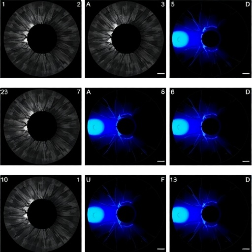
Image Credits: AI Generated
DOI: 10.1007/s44163-025-00795-8
Keywords: Retina, Optic Disc Segmentation, U-Net Architecture, Medical Imaging, Artificial Intelligence, Fundus Images, Deep Learning.
Tags: advancements in image analysis technologyautomated optic disc segmentationcustomized deep learning models for healthcarediabetic retinopathy diagnosis toolsenhancing diagnostic accuracy in ophthalmologyglaucoma detection using AIimproving consistency in retinal disease diagnosismachine learning in retinal disease assessmentminimizing human error in medical imagingoptimizing neural networks for medical applicationsretinal fundus image analysisU-Net architecture for retinal segmentation


