Complex field imaging provides a detailed structural analysis of samples by capturing both the amplitude and phase information of objects, offering insights into properties like absorption and refractive index distributions that reveal the sample’s morphology and composition. However, conventional sensors typically capture only the amplitude information of the specimen, resulting in the loss of crucial phase information unless complex interferometric or holographic methods are utilized.
Credit: by Jingxi Li, Yuhang Li, Tianyi Gan, Che-Yung Shen, Mona Jarrahi, and Aydogan Ozcan
Complex field imaging provides a detailed structural analysis of samples by capturing both the amplitude and phase information of objects, offering insights into properties like absorption and refractive index distributions that reveal the sample’s morphology and composition. However, conventional sensors typically capture only the amplitude information of the specimen, resulting in the loss of crucial phase information unless complex interferometric or holographic methods are utilized.
A team of UCLA researchers introduced an all-optical complex field imaging technique that directly captures both the amplitude and phase information of optical fields using an intensity-based sensor array. This innovative approach, detailed in their publication in Light: Science & Applications, marks a significant departure from traditional methods, which often involve complex hardware and high computational load, often using iterative algorithms.
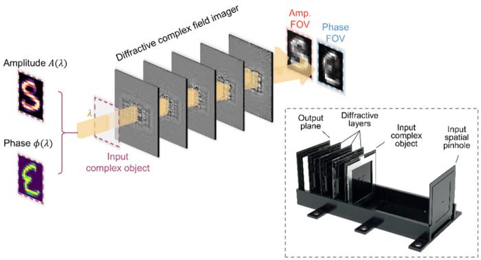
This breakthrough is grounded in the design of a complex field imager capable of directly capturing both the amplitude and phase distributions of an incoming light using an array of intensity-only detectors. As detailed in their research, the device incorporates multiple diffractive surfaces—each designed and optimized through supervised deep learning algorithms—to facilitate precise spatial modulation of incoming complex fields. This diffractive architecture, also known as a diffractive deep neural network (D2NN), has previously demonstrated its utility in a variety of all-optical information processing tasks, including image classification, encryption, spectral encoding, and visual computing, among others.
This diffractive complex-field imaging technology effectively maps the amplitude of the incoming light onto a specific output field dedicated to amplitude imaging while simultaneously converting the wave’s phase information into an intensity pattern used for quantitative phase imaging. For any arbitrary complex object or field at the device’s input, the resulting intensity distributions directly reveal the sample’s amplitude and phase information through a single image capture step.
“By using deep learning-optimized diffractive surfaces, we created a physical system that directly captures the complex field of an input object, eliminating the need for traditional digital reconstruction algorithms,” explained Dr. Aydogan Ozcan, the project’s principal investigator and the Chancellor’s Professor at UCLA. “This makes the imaging process much simpler and faster, reducing the hardware footprint and energy consumption significantly.”
Furthermore, the team explored various other designs that incorporate wavelength multiplexing to capture amplitude and phase information of samples at different wavelengths. Such a design has been validated through 3D-printed prototypes that operate in the terahertz spectrum. The experimental outcomes from these prototypes closely align with the numerical simulations, confirming their potential for practical applications.
“We envision this technology having broad implications across several fields, including security, biomedical imaging, and material science. Its ability to perform rapid and accurate imaging without digital image reconstruction algorithms could redefine standard practices in these areas,” Dr. Ozcan added.
UCLA researchers emphasize that this technology not only demonstrates the feasibility of all-optical complex field imaging but also highlights its potential to be adapted across various wavelengths of the electromagnetic spectrum, including the visible and infrared spectra, without the need to retrain or redesign the optimized diffractive layers.
Joining Dr. Ozcan in this pioneering work are the graduate students Jingxi Li, Yuhang Li, Tianyi Gan, Che-Yung Shen, as well as Prof. Mona Jarrahi, all from the Electrical and Computer Engineering department at UCLA. Dr. Ozcan holds faculty appointments in the bioengineering department and serves as an associate director of the California NanoSystems Institute (CNSI).
Journal
Light Science & Applications
DOI
10.1038/s41377-024-01482-6


