New Insights into How Prenatal Oxycodone Exposure Alters Placental Communication and Heightens Risks for Fetal Heart Disease
Emerging evidence from a groundbreaking experimental study unravels the complex molecular dialogue between the placenta and developing fetus disrupted by maternal oxycodone use during pregnancy. Published in the journal Extracellular Vesicles and Circulating Nucleic Acids, this research elucidates how chronic in utero exposure to oxycodone profoundly reshapes placental small extracellular vesicles (sEVs), at both a structural and proteomic level, potentially setting the stage for increased fetal cardiac vulnerability.
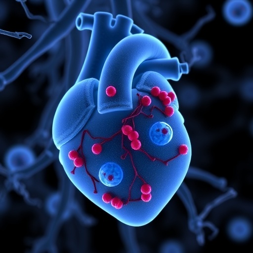
The placenta, a transient yet pivotal organ, governs fetal development by mediating nutrient exchange and transmitting vital biochemical signals. Among these signaling vehicles are sEVs—nanometer-scale, membrane-bound vesicles that ferry proteins, nucleic acids, and lipids between maternal and fetal compartments. These vesicles effectively act as molecular messengers, influencing diverse fetal developmental processes, especially the intricate progression of cardiac formation and function.
Utilizing advanced electron microscopy and nanoparticle tracking analysis, the research team meticulously compared sEV populations isolated from oxycodone-exposed pregnancies against saline-treated controls. Notably, oxycodone exposure led to a significant increase in the quantity of sEVs while concurrently reducing their average size. This alteration hints at stress-induced modifications in vesicle biogenesis and cargo packaging mechanisms—hallmarks of a placenta undergoing adaptive or maladaptive responses to opioid insult.
Perhaps most strikingly, a comprehensive proteomic survey identified 456 distinct proteins within the placental sEVs, of which over 100 exhibited significant differential expression attributable to oxycodone exposure. Proteins involved in fundamental cellular processes such as protein synthesis and vesicular transport were upregulated, suggesting an attempt by the placenta to reorganize its intracellular trafficking systems under duress. Conversely, a profound downregulation was observed in proteins key to metabolic pathways, including fatty acid catabolism, mitochondrial energy production, and detoxification enzymes, underscoring a compromised metabolic capacity and weakened support for the developing fetus.
Among the suppressed proteins, five critical molecules—Atp2a2, Lmna, Tgfb3, Agt, and Sgce—stand out for their established links to cardiomyopathic conditions. Atp2a2 encodes a calcium pump regulating cardiac contractility; Lmna is essential for nuclear envelope integrity; Tgfb3 modulates cardiac remodeling; Agt participates in the renin-angiotensin system influencing blood pressure; and Sgce relates to muscle function. Their coordinated downregulation proposes a mechanistic pathway where altered placental sEV cargo directly impairs heart muscle integrity in the fetus, predisposing to cardiomyopathy.
These findings highlight how the placenta’s maladaptive response to sustained opioid exposure extends beyond mere nutrient transport dysfunction, advancing into the realm of targeted molecular miscommunication. By modifying the proteome of sEVs, the placenta inadvertently transmits pathogenic signals that could disrupt fetal cardiac developmental trajectories and prime offspring for future cardiovascular morbidity.
Importantly, the detection of these protein alterations in sEVs introduces a promising avenue for noninvasive prenatal biomarker development. Tracking placental sEV signatures in maternal blood samples could enable early identification of opioid-exposed pregnancies at elevated risk for fetal cardiac anomalies. Such biomarkers would be invaluable for timely clinical interventions aimed at mitigating adverse outcomes in newborns affected by in utero opioid exposure.
Moreover, this study establishes a robust preclinical model to explore therapeutic strategies that may restore normal placental sEV composition or block detrimental signaling pathways. Interventions targeting sEV biogenesis or cargo selection hold potential to shield fetal hearts from opioid-induced injury, fostering healthier pregnancy outcomes amidst the ongoing opioid crisis.
The use of proteomics combined with high-resolution imaging underscores the power of integrative, systems-level analyses to decode the subtle yet consequential effects of maternal drug use on fetal development. This multi-modal approach reveals how chronic oxycodone modifies not just individual proteins but entire molecular networks orchestrating placental-fetal communication.
Altogether, this pioneering research enriches the scientific understanding of how prenatal opioid exposure compromises fetal health through altered vesicle-mediated intercellular messaging. It calls for heightened awareness of opioid impacts during pregnancy and bolsters the rationale for developing biomarker-driven monitoring and targeted therapies to safeguard cardiac development.
As opioid use continues to rise globally, unraveling the molecular underpinnings of its effects on placental function and fetal organogenesis is critical. This study paves the way for future translational research that can transform obstetric and neonatal care for opioid-exposed populations, ultimately reducing the burden of congenital heart defects linked to prenatal drug exposure.
The comprehensive analysis of placental sEV proteome alterations and their relationship with fetal cardiomyopathy-linked pathways represents a significant leap forward in maternal-fetal medicine. It spotlights the placenta not only as a barrier and conduit but also as a dynamic signaling hub whose integrity is vital for the lifelong cardiovascular health of the offspring.
Subject of Research: Not applicable
Article Title: Chronic in utero oxycodone exposure alters placental small EV proteome and fetal cardiomyopathy-linked pathways
News Publication Date: 10-Feb-2026
Web References: http://dx.doi.org/10.20517/evcna.2025.138
Image Credits: HIGHER EDUCATION PRESS
Keywords: Cell biology, placental small extracellular vesicles, oxycodone exposure, fetal cardiomyopathy, proteomics, placenta-fetus communication, prenatal opioid effects, biomarker discovery
Tags: electron microscopy of placental vesiclesfetal cardiac vulnerability from opioidsfetal heart stress biomarkersin utero oxycodone exposure risksmaternal drug use and fetal developmentmolecular communication in placentananoparticle tracking in pregnancy researchopioid exposure during pregnancyplacental signaling and fetal heart diseaseplacental small extracellular vesiclesprenatal oxycodone effects on placentaproteomic changes in placental vesicles

