A pioneering feasibility investigation has utilized open-field magnetic resonance imaging to record the dynamic biomechanical adaptations of the maternal pelvis and pelvic floor during the second stage of active labor in real time. Published in European Radiology Experimental, the proof-of-concept study demonstrates that advanced intrapartum imaging can directly capture the complex physical deformations of maternal soft tissues, musculature, and bony articulations as the fetal head negotiates the birth canal. Clinical understanding of intrapartum mechanics has historically been constrained by the opacity of the pelvic cavity and the logistical obstacles associated with conducting advanced radiological examinations during active childbirth. By employing an open-geometry scanner situated adjacent to a maternity ward, investigators visualized how the muscular pelvic floor stretches, inverts, and shifts under mechanical pressure, offering an unprecedented descriptive window into human parturition.
For decades, obstetric assessments during labor have depended almost entirely on manual digital vaginal examinations, a subjective technique that relies solely on palpation of accessible maternal structures and fetal landmarks. Although transperineal ultrasound has refined the evaluation of fetal head station relative to the pubic symphysis, its acoustic window is frequently impeded by dense pelvic bones and fetal acoustic shadowing. Crucial internal landmarks, including maternal ischial spines, the precise flexion attitude of the fetal head, and deep fascial planes of the pelvic diaphragm, remain largely obscured during sonography. Biomechanical models and computer simulations have attempted to recreate these complex spatial dynamics, but they have lacked empirical, real-time confirmation in living patients. By capitalizing on magnetic resonance imaging, which visualizes osseous and soft-tissue elements across multiple planes without ionizing radiation, the research team sought to bridge the persistent gap between computational predictions and physiological reality.
Conducted under the clinical protocol designated as the IMAGINAITRE study, the single-center prospective pilot investigation received official authorization from the French National Agency for Drug and Medical Product Safety and ethical approval from the regional Institutional Review Board. Recruitment focused on healthy pregnant women aged between twenty-three and thirty-four years carrying singleton pregnancies at thirty-nine weeks of gestation or greater with confirmed cephalic presentation. Candidates presenting with obstetric complications, non-cephalic presentations, prior uterine surgical scarring, multiple gestations, or magnetic resonance imaging contraindications were excluded. A total of twenty-seven eligible women consented and enrolled in the research program. Each volunteer was scheduled to undergo an initial baseline scan within two hours before labor onset, followed by a second intrapartum scanning session upon reaching the second stage of labor, defined by complete cervical dilation and engagement of the fetal head past the superior pelvic strait.
Logistical constraints and the unpredictable tempo of labor prevented twenty participants from completing the intrapartum imaging phase, leaving a cohort of seven patients who successfully underwent both pre-labor and active intrapartum examinations. These seven women had a mean age of twenty-eight years and exhibited adequate pelvic capacity, as evidenced by Magnin pelvic scores exceeding twenty-five points, with a median score of 26.1 across a range from 25.1 to 27.5. Three participants were primiparous, three were secundiparous, and one was tertiparous. Five achieved uncomplicated spontaneous vaginal deliveries, whereas two post-term participants underwent unplanned Cesarean deliveries due to arrest of descent at full dilation. Neonatal birth weights ranged from 3,095 grams to 4,525 grams, with a median of 3,755 grams. No maternal postpartum hemorrhages, instances of shoulder dystocia, or neonatal depressions occurred, confirming that imaging did not adversely impact clinical care.
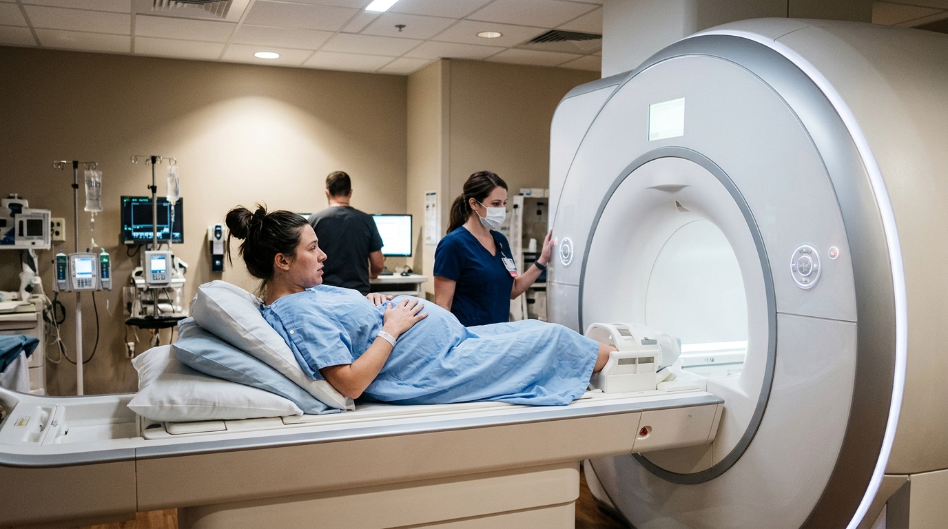
Executing intrapartum imaging safely required an open 1-Tesla Philips Panorama scanner located directly within the maternity facility, allowing rapid transit between clinical suites. Patient transfer from the delivery room to the scanner took a median of 2.6 minutes, ranging from 2.4 to 3.2 minutes, while transfer from the scanner to surgery for unplanned Cesarean deliveries averaged 2.5 minutes. Maternal positioning was maintained in a modified supine posture mirroring delivery bed ergonomics. The protocol avoided intravenous contrast agents and utilized rapid sequences, including multiplanar T1-weighted turbo field echo images, three-dimensional isotropic acquisitions, and dynamic mid-sagittal sequences lasting approximately sixty seconds. Total active scanning time averaged eight minutes, with no continuous sequence exceeding two minutes. Fetal heart rate surveillance was briefly paused for cumulative intervals never exceeding ten minutes per patient, with multidisciplinary obstetrical, neonatological, and radiological teams present to maintain continuous oversight.
Detailed image analysis revealed profound geometric rearrangements across the maternal muscular floor as the fetal head advanced past the pelvic inlet. Before labor onset, the levator ani muscle group, especially its iliococcygeal portion, presented as a supportive, upwardly convex hammock spanning beneath the pelvic viscera. During intrapartum descent, direct compressive loading from the fetal cranium completely flattened this muscular architecture, forcing it into an inverted, concave configuration. This inversion was accompanied by substantial enlargement of the urogenital hiatus and severe stretching of the puborectal fascia. Surrounding pelvic fat and connective tissue elements, particularly the adipose reserves located between the vaginal wall and the sacrotuberous ligaments, shifted inferiorly and paramedially into zones of lesser mechanical resistance. This soft-tissue displacement occurred predominantly toward the anal sphincter region, culminating in the prominent perineal bulge recognized during active delivery.
Bony structures exhibited distinct directional movements strictly confined to the sagittal plane without lateral pelvic widening. In all seven participants, fetal descent induced marked posterior deflection of the coccyx via nutation of the sacrococcygeal articulation. Quantitative morphometry documented that the distance from the inferior margin of the pubic symphysis to the coccyx increased by an average of 0.78 centimeters, ranging between 0.48 and 1.35 centimeters. Concurrently, the distance from the coccyx to the sacral promontory expanded by a mean of 0.80 centimeters, with measured values spanning from 0.76 to 1.25 centimeters. As a consequence of this sacrococcygeal tilting, the interdiameter angle between these two reference lines decreased by an average of 5.3 degrees. Importantly, measurements of the pelvic inlet, mid-pelvis, and outlet demonstrated that the bony pelvis behaved as a rigid ring, confirming that birth canal expansion occurred entirely through soft-tissue distension.
To quantify three-dimensional displacements within the internal fascial scaffolding, the investigators segmented the tendinous arch of the levator ani using open-source modeling software. By co-registering pre-labor and intrapartum scans to static pelvic reference landmarks, including the acetabula and pelvic brim, the team tracked structural deviations across seven standardized measurement stations. The tendinous arch, which originates along the medial fascia of the obturator internus muscle and anchors the iliococcygeus, exhibited substantial inferior and posterior displacement under the mechanical strain of fetal progression. The magnitude of this caudal excursion was unevenly distributed along its anatomical length, displaying maximal downward displacement along its anterior segment and progressively diminishing excursions through its medial and posterior segments. Furthermore, transverse spacing between the bilateral tendinous arches remained essentially unchanged, confirming that fascial loading was primarily absorbed in the vertical and sagittal planes rather than through lateral spreading.
The soft tissues surrounding the perineal body and anal sphincter complex experienced the most extreme mechanical distension recorded during the study. As the fetal head descended toward crowning, the midline perineal raphe rotated dramatically from a horizontal orientation into an upright vertical alignment while being thrust posteriorly. Simultaneously, the puborectalis muscle fibers separated laterally and the anal canal underwent noticeable radial dilation. In three participants where precise anatomical tracking was feasible, the longitudinal length of the anal sphincter increased by an average of 1.0 centimeter, representing a proportional elongation of 27.9 percent across a range of 10.7 to 47.0 percent. Transverse sphincter dimension increased by an average of 0.9 centimeters, demonstrating an average widening of 31.3 percent, though one patient exhibited localized thinning. Data indicated that deeper fetal head descent correlated with decreased residual sphincter width, highlighting severe tensile forces underlying postpartum pelvic trauma.
Unexpected anatomical behavior was observed regarding the maternal urinary bladder during the second stage. In standard obstetric management, clinicians frequently catheterize or empty the bladder, operating on the assumption that luminal fluid volume obstructs fetal progression through the birth canal. However, intrapartum scans demonstrated that six of the seven patients possessed a partially filled bladder, which was displaced superiorly above the pubic symphysis as the fetal head engaged. This upward migration relocated the organ out of the direct line of downward force, shielding it from direct cranial compression. Conversely, in the single participant who possessed a completely empty bladder, the collapsed bladder walls were pinned between the descending fetal skull and the pubic bone, subjecting the organ to direct shearing forces. Fetal head descent distances and progression angles were comparable regardless of bladder filling, generating the hypothesis that maintaining some bladder volume might protect maternal tissues.
Despite providing exceptional structural insight, the investigators emphasize that findings from this proof-of-concept study remain strictly preliminary, descriptive, and hypothesis-generating. The small cohort of seven completing patients reflects the profound logistical difficulty of coordinating advanced intrapartum imaging without compromising maternal or neonatal safety. The study was not designed or powered for statistical hypothesis testing, and opportunistic scan timing captured only isolated intervals of the delivery process rather than the entire continuum. Furthermore, the absence of concurrent comparative modalities such as transperineal ultrasound prevents direct cross-validation of measurements against routine clinical monitoring tools. The researchers caution that these initial observations must not be interpreted as justification for altering clinical guidelines or intrapartum protocols. Instead, this feasibility demonstration lays essential methodological groundwork, showing that intrapartum open magnetic resonance imaging is viable and opening realistic pathways for multicenter investigations into pelvic floor protection during childbirth.
Subject of Research: Feasibility of open-field intrapartum magnetic resonance imaging for evaluating maternal pelvic biomechanics during labor
Article Title: MRI visualization of maternal pelvic and perineal anatomical changes during the second stage of labor: a feasibility study
Article References: Maran, J.-C., Ami, O., Dao, T. T., Mage, G., & Boyer, L. (2026). MRI visualization of maternal pelvic and perineal anatomical changes during the second stage of labor: a feasibility study. European Radiology Experimental, 10(1), Article 129. https://doi.org/10.1186/s41747-026-00757-4
Image Credits: AI Generated
DOI: 10.1186/s41747-026-00757-4
Keywords: intrapartum MRI, second stage of labor, pelvic floor biomechanics, levator ani muscle, coccygeal nutation, perineal expansion, anal sphincter stretching, open-field MRI, fetal head descent, maternal anatomy, pelvic outlet dimensions, urinary bladder displacement, tendinous arch of levator ani, obstetric imaging, cervical effacement, birth canal mechanics, pudendal anatomy
News Source: Ophelia Keating. (November 16, 2026). Open MRI Reveals Real-Time Maternal Pelvic Adaptations During Active Labor. Scienmag.