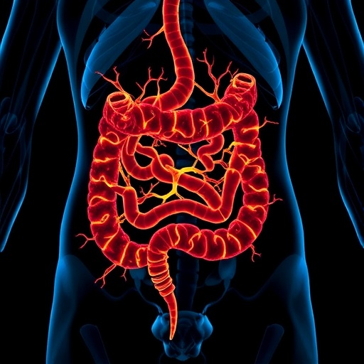
A groundbreaking study published in Nature reveals new insights into how nutrients activate specific networks of enteric neurons in the small intestine, deepening our understanding of gut neurobiology and its intricate communication pathways. This research uncovers the mechanisms by which distinct neuronal populations within the enteric nervous system (ENS) respond to localized stimuli, shedding light on the direct and synaptic activation processes of intrinsic primary afferent neurons (IPANs) and other neuronal subtypes.
.adsslot_onMS5utR6b{ width:728px !important; height:90px !important; }
@media (max-width:1199px) { .adsslot_onMS5utR6b{ width:468px !important; height:60px !important; } }
@media (max-width:767px) { .adsslot_onMS5utR6b{ width:320px !important; height:50px !important; } }
ADVERTISEMENT
To further delineate the functional connectivity between individual intestinal villi and the myenteric neural network, the investigation utilized focal electrical stimulation at 20 Hz for one second directed at a single villus. This localized stimulation elicited reproducible calcium transients in approximately 14% of myenteric neurons within the imaging field, forming a consistent responder population. Immunohistochemical analyses further linked these electrical responders predominantly to calbindin-expressing neurons, reinforcing their identity as IPANs or related sensory neurons. These observations highlight the precision of enteric neuronal recruitment orchestrated at the level of single mucosal structures.
Under hexamethonium treatment during electrical stimulation, although the total number of initially activated neurons remained similar, a slight reduction in response amplitude was observed, indicating that while direct stimulation predominates, a subset of synaptically mediated responses contributes to the overall activity pattern. This nuanced interplay between direct and synaptic activation underscores the complexity of enteric neuronal communication and may reflect diverse physiological roles—ranging from sensory input to coordinated motor responses.
The involvement of serotonergic (5-HT) signaling was also implicated through comparison with intravillus injections of 5-HT, which yielded a similarly sized population of responsive myenteric neurons. This correlation suggests a broad sensitivity of mucosal nerve endings to serotonin, a critical neurotransmitter and modulator within the ENS, known for regulating motility, secretion, and local reflexes. This finding bridges chemical neuromodulation and electrical activation in sensory enteric neurons connected to mucosal structures.
Simultaneous imaging of calcium transients in both the myenteric and submucosal plexuses revealed temporal dynamics whereby myenteric neuronal responses precede submucosal ones. This sequential activation pattern may reflect hierarchical processing of sensory inputs and subsequent modulation of secretomotor activities, supporting the concept of layered reflex circuits within the gut wall. Such insights enhance our understanding of how the ENS integrates chemical and electrical information to regulate intestinal function.
The study’s methodological advances, including the use of spinning disk confocal microscopy and genetically identified neuronal markers, enabled unprecedented resolution in mapping functional connectivity between neurons and mucosal elements in the small intestine. This approach allowed the authors to distinguish direct responses from those mediated by synaptic transmission with pharmacological tools, setting a new standard for experimental exploration of enteric neural circuits.
Moreover, the observation that removing the myenteric plexus attenuated submucosal neuron responses to villus stimulation confirms inter-plexus communication, highlighting the myenteric plexus’s critical role in modulating network activity. This crosstalk may be essential for coordinating complex intestinal reflexes involving motility and secretion, emphasizing the enteric nervous system’s decentralized but integrated nature.
These findings offer compelling evidence that the ENS’s neuronal complexity extends beyond its anatomical layering to functional specialization and connectivity finely tuned to local stimuli. The identification of distinct neuronal responder populations tied to mucosal activation underscores the possibility that different nutrient signals could selectively recruit specific enteric circuits, influencing digestive processes and gut homeostasis.
In sum, this comprehensive analysis of enteric neuronal activation dynamics provides a deeper mechanistic understanding of how the gut senses and responds to its chemical environment. It opens avenues for targeted interventions in disorders of gastrointestinal motility and sensitivity, such as irritable bowel syndrome (IBS) and other functional bowel disorders, where enteric neuronal dysfunction is implicated.
By revealing how nutrients and localized stimuli engage discrete neuronal networks within the small intestine, this study not only advances fundamental gastrointestinal neuroscience but also sets a foundation for novel therapeutic strategies that harness the ENS’s intrinsic capabilities. This research elevates the enteric nervous system from a simple relay station to an intricate sensory organ capable of precise and adaptable neural processing.
As science continues to unravel the complexities of the gut-brain axis, studies like this emphasize the ENS’s pivotal role, demonstrating that the gut itself contains neural circuits with sophisticated integrative properties. Understanding these circuits brings us closer to manipulating gut sensory pathways to improve health outcomes beyond traditional pharmacology.
This study truly exemplifies a landmark in enteric neuroscience, combining state-of-the-art imaging, pharmacology, and molecular markers to decode the language of the gut’s neural networks. Future research may explore how these neuronal activation patterns vary in disease states or adapt to different dietary inputs, further enriching our knowledge of intestinal physiology and pathophysiology.
Subject of Research: Enteric nervous system neuronal activation and connectivity in the small intestine
Article Title: Nutrients activate distinct patterns of small-intestinal enteric neurons
Article References:
Fung, C., Venneman, T., Holland, A.M. et al. Nutrients activate distinct patterns of small-intestinal enteric neurons. Nature (2025). https://doi.org/10.1038/s41586-025-09228-z
Image Credits: AI Generated
Tags: calbindin-positive myenteric neuronsdirect neuronal activation processesenteric nervous system mechanismsenteric neuron population responsesgut neurobiology insightshigh potassium stimulation effectsintrinsic primary afferent neurons activationmucosal stimulation researchneurophysiology of the gutneurotransmission in the small intestinesmall intestine neuron patternssynaptic transmission in ENS


