A multicenter randomized clinical trial alongside in vitro degradation testing has demonstrated that a novel cranial flap fixation system manufactured entirely from high-purity poly-L-lactic acid can securely stabilize cranial bone flaps following craniotomy while avoiding the structural limitations of existing resorbable clamps. Cranial bone flap repositioning represents a fundamental concluding stage in craniotomy procedures, yet conventional closure methods have historically introduced compromises between rigid mechanical stability, biological compatibility, and complete long-term tissue integration. By addressing persistent material and mechanical challenges identified in older generation clamps, the trial findings provide rigorous clinical and laboratory validation for an entirely absorbable, structurally adjustable alternative that maintains necessary mechanical resistance through the biological bone healing window before slowly degrading without triggering localized adverse events.
Neurosurgical craniotomies necessitate the temporary removal of a section of skull bone to expose the underlying dura mater and brain tissue during operations for cranial trauma, intracranial aneurysms, and space-occupying tumors such as meningiomas. Following intracranial intervention, stable surgical fixation of the retracted bone flap is essential to restore structural skull integrity, shield vulnerable cerebral tissues from trauma, prevent cosmetic skull deformities, and avoid severe postoperative complications such as cerebrospinal fluid leakage, meningoencephalocele, and bone flap resorption. The historical evolution of bone flap stabilization has progressed from manual suturing and stainless steel wires to single-sided metal plates and specialized dual-sided clamping systems. Early techniques involving surgical sutures suffered from inherent instability, frequent loosening, and suture rupture that led directly to fixation failure. Stainless steel wire offered improved rigidity but required cumbersome operative handling, while the resulting wire knots frequently irritated overlying scalp tissue, eroded cutaneous layers, and produced hardware exposure. While modern fixation plates provide high structural toughness and remain common in routine practice, their single-sided application leaves them vulnerable to external compressive forces that can depress the bone flap inward toward intracranial structures.
Dual-sided fixation clamps circumvent many of these traditional vulnerabilities by securing the free bone flap against the surrounding cranial margin through paired internal and external discs. This bilateral clamping mechanism relies on upper and lower discs that sandwich the bone margins, producing consistent clamping force without requiring bone screws that penetrate the inner cortical table of the skull. The absence of penetrating screws eliminates the danger of hardware advancing into the intracranial space or piercing the underlying dura mater, making clamping devices especially advantageous in thinner pediatric skulls where standard screw purchase is limited or poses developmental hazards. Because the human skull primarily experiences longitudinal mechanical pressure, the bilateral distribution of force delivered by dual-sided clamps establishes superior mechanical stability compared to single-sided plates. This configuration maintains smooth structural contact between the free bone flap and the margins of the surgical bone window, preserving an anatomical barrier that actively encourages osteogenic bridging and uneventful biological bone fusion.
Despite the functional advantages offered by bilateral clamping, the material composition of implantable devices introduces distinct clinical considerations. Permanent fixation systems fabricated from titanium alloy provide exceptional mechanical resilience and wear resistance, yet they cause significant artifacts on subsequent computed tomography and magnetic resonance imaging scans while slowly releasing trace metallic ions over extended periods. Polyether ether ketone has emerged as an alternative nonabsorbable polymer that reduces radiographic artifacts, but it exhibits lower mechanical strength than metals, carries risks of long-term structural wear, and still requires permanent anchoring screws that may disrupt subsequent pediatric skull growth. Absorbable fixation systems overcome these permanent hardware drawbacks by offering biodegradability, radiographic translucency, non-paramagnetism, and an absence of magnetothermal heating during diagnostic neuroimaging. However, widely used commercial devices such as the Aesculap CranioFix absorbable clamp present documented practical drawbacks, including incomplete long-term bodily absorption, a rigid disc orientation that cannot adapt to the natural curvature of the skull, and an inability to loosen or readjust the clamp once the connecting elements are locked in place.
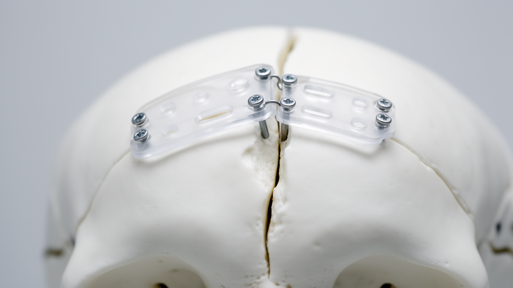
To overcome these functional and material limitations, researchers developed a novel absorbable system constructed exclusively from high-purity, semicrystalline poly-L-lactic acid designed to degrade fully into benign L-lactic acid metabolites. The mechanical architecture incorporates five distinct components consisting of an upper column lock, a lower column lock, a connecting rod, an upper disc, and an articulated lower disc. The column lock mechanism features a calibrated, fixed-force column design that delivers a uniform preload during surgical tightening, eliminating manual variation in clamping pressure. Furthermore, before final lock disengagement, the device permits surgical readjustment: operating surgeons can reverse the rotation of the lower column lock to loosen the upper disc, reposition the clamp relative to the cranial margins, and retighten the assembly without damaging components or having to cut and discard the implant. The central connecting rod features a flattened, threaded profile that fits into narrow osteotomy kerfs, enlarges the contact surface area against the bone window margin to prevent horizontal slippage, and terminates in a hanging platform that grants angular mobility to the lower disc so it conforms smoothly to the internal contour of the inner skull table.
Laboratory evaluation began with in vitro accelerated degradation testing conducted in phosphate buffer solution adjusted to a physiological pH of 7.4 and maintained at seventy degrees Celsius to compare the material stability, mechanical endurance, and degradation profiles of both systems. In these accelerated bench tests, the experimental poly-L-lactic acid device required fourteen days for its intrinsic viscosity to decline below the defined testing endpoint threshold of 0.1 deciliters per gram, whereas the control CranioFix material reached this endpoint in only seven days. Mechanical evaluation measuring tripping force demonstrated that the experimental device maintained superior resistance against displacement throughout the accelerated testing period. Although both systems experienced sharp declines in retention force by the third day of accelerated exposure, the control clamp dropped below the recognized clinical safety fixation threshold of thirty Newtons on day three, whereas the experimental clamp maintained force exceeding this threshold until day four. Analysis of degradation chemistry revealed that the control device released an initial burst of lactic acid between days three and seven, averaging a release rate of 40.09 milligrams per day, whereas the experimental material exhibited delayed, steady release at 15.42 milligrams per day after day seven, reflecting its higher initial polymer crystallinity.
Following in vitro verification, the research team conducted a multicenter, randomized, parallel-group, positive-controlled non-inferiority clinical trial spanning four major tertiary medical centers in China: Xiangya Hospital of Central South University, Renmin Hospital of Wuhan University, the First Hospital of Hunan University of Chinese Medicine, and West China Hospital of Sichuan University. Between September 4, 2017, and July 8, 2019, investigators screened ninety-four adult patients requiring cranial flap fixation after craniotomy for nonmetastatic intracranial lesions or non-infected cranial trauma. Ninety eligible subjects aged eighteen to sixty-nine years were randomized into experimental and control cohorts, with eighty-seven patients ultimately undergoing surgery and receiving the assigned fixation device. Patients with underlying metabolic or cardiovascular disorders, local scalp infections, lactic acid allergies, or planned surgical bone flap decompression were excluded from enrollment to isolate the biological and mechanical performance of the implants. The trial was formally registered with the Chinese Clinical Trial Registry under identifier ChiCTR2500099674.
Clinical efficacy was systematically evaluated across a forty-eight-week postoperative follow-up protocol utilizing three-dimensional computed tomography reconstructions generated using specialized visualization software to track cranial flap positioning, osteotomy gap dimensions, and implant resorption kinetics. Quantitative imaging demonstrated that the mean bone gap distance decreased progressively over time in both cohorts as physiological synostosis took place. In the experimental cohort, the mean bone gap narrowed from 1.61 millimeters at week one to 1.00 millimeter at week twelve, 0.79 millimeters at week twenty-four, and 0.58 millimeters at week forty-eight. In the control cohort, the corresponding gap measurements were 1.51 millimeters at week one, 1.17 millimeters at week twelve, 1.03 millimeters at week twenty-four, and 0.80 millimeters at week forty-eight.
The secondary clinical outcome evaluating cranial stability demonstrated exceptional performance for the novel design, with the experimental cohort achieving a one hundred percent qualified rate for bone flap displacement across all postoperative assessment intervals. Surgical displacement of less than two millimeters was defined a priori as clinically qualified. In contrast, the control group exhibited one unqualified case of bone flap displacement exceeding two millimeters at one week postoperatively, representing a qualified rate of 97.78 percent, and two unqualified cases at weeks six, twelve, twenty-four, and forty-eight, yielding a qualified rate of 95.56 percent. Three-dimensional volumetric tracking of the polymer implants indicated distinct in vivo resorption profiles between the two materials. The experimental poly-L-lactic acid clamps resisted early volumetric loss, retaining approximately 63.6 percent of their original structural volume at twenty-four weeks and 48.8 percent at forty-eight weeks. Conversely, control implants exhibited noticeable structural degradation by postoperative week six, declining to 54.6 percent of original volume, and dropping below half of their starting volume to 43.9 percent by week twelve.
Because clinical skull consolidation typically requires twelve to twenty-four weeks, with final synostosis frequently extending to forty-eight weeks or longer, retaining clamp integrity throughout this window ensures that external mechanical forces do not displace healing bone margins. The rapid early volume loss observed in the control group reflected the amorphous nature of its poly(L-lactide-co-D,L-lactide) copolymer, which degraded rapidly but structurally failed before bone bridging was fully finalized. Furthermore, the control system incorporated permanent braided polyester sutures to connect its resorbable discs, leaving unabsorbed foreign material permanently embedded within cranial tissues. In contrast, the experimental system utilized solely pure poly-L-lactic acid without nonresorbable sutures or metal elements, permitting complete, uniform biodegradation without leaving permanent synthetic residues that could incite late foreign-body reactions or restrict regional tissue remodeling.
Safety assessments encompassing routine hematological indices, hepatic and renal biochemical panels, and systemic coagulation profiles revealed no statistically significant differences between the two treatment groups when comparing baseline preoperative laboratory values to tests obtained one week after surgery. Postoperative clinical monitoring recorded common neurosurgical complications across both cohorts, including mild local wound infections, epidural effusions, and subcutaneous fluid collections, all of which resolved following standard symptomatic care without necessitating surgical revision. The lower disc of the experimental device incorporates specialized perforations designed to promote fluid drainage, effectively mitigating dead-space fluid pooling beneath the hardware. Across the entire clinical evaluation, no device-related adverse events were documented in the experimental cohort. In the control cohort, one patient experienced an adverse event deemed possibly related to the implant, manifesting as poor local incision healing that subsequently resolved under appropriate clinical management, representing an adverse incidence rate of 2.22 percent.
While these comprehensive in vitro and clinical findings demonstrate substantial advantages for the novel resorbable fixation system, the authors noted specific methodological boundaries governing the interpretation of their data. The clinical trial deliberately restricted participant eligibility to adult patients aged eighteen to seventy years to ensure patient safety and uniform biological responses during initial evaluation, excluding younger pediatric cohorts who stand to benefit substantially from completely absorbable cranial hardware that accommodates ongoing brain and skull expansion. Nevertheless, by combining an adjustable, force-limiting bilateral locking mechanism with a biocompatible, slow-degrading poly-L-lactic acid matrix, the investigated system successfully addresses the operational and material deficiencies of earlier resorbable clamps, delivering reliable mechanical fixation and favorable safety in modern cranial flap surgery.
Subject of Research: Comparative in vitro and clinical evaluation of an absorbable cranial flap fixation system versus Aesculap CranioFix
Article Title: A comparative study of a novel absorbable cranial flap fixation system and Aesculap CranioFix
Article References: Teng, C., Liang, H., Yang, Q., Fang, Y., Jian, Z., Yu, J., Luo, G., Han, X., Du, J., Wanggou, S., & Li, X. (2025). A comparative study of a novel absorbable cranial flap fixation system and Aesculap CranioFix. Chinese Neurosurgical Journal, 11(1), Article 18. https://doi.org/10.1186/s41016-025-00406-6
Image Credits: AI Generated
DOI: 10.1186/s41016-025-00406-6
Keywords: craniotomy, bone flap fixation, absorbable implants, poly-L-lactic acid, Aesculap CranioFix, cranial reconstruction, clinical trial, neurosurgery, biodegradable polymer, bone gap healing, three-dimensional CT, biocompatibility, implant degradation, mechanical stability, surgical clamps, tripping force, intrinsic viscosity
News Source: Ophelia Keating. (November 14, 2026). Novel Resorbable Skull Flap Fixation System Matches Standard Device Performance in Trial. Scienmag.