
Credit: N Narayan et al. 2018, University of Oxford, Headington, Oxford, UK
RESTON, VA – A new positron emission tomography (PET) imaging method more fully evaluates the extent of rheumatoid arthritis by targeting translocator protein (TSPO) expression in the synovium (joint lining tissue). The study is featured in the July issue of The Journal of Nuclear Medicine.
PET radioligands that target TSPO, which is highly expressed on activated macrophages (immune cells responding to inflammation), have proved an excellent tool for imaging joint inflammation in rheumatoid arthritis (RA).
"TSPO-targeted imaging has long been used as a means of imaging macrophage infiltration in vivo," explains Nehal Narayan of the University of Oxford in Oxford, U.K. "Numerous studies have demonstrated TSPO-targeted PET as a highly sensitive and specific means of imaging synovitis [inflammation of joint lining tissue], purportedly through imaging synovial macrophage infiltration, a critical process in RA pathogenesis. However, this premise does not take into account the ubiquitous expression of TSPO."
She points out, "Here we present the first ever analysis of TSPO expression in the major constituents of RA pannus (inflamed synovium), demonstrating that TSPO PET likely acts as an imaging tool of not only macrophages, but also activated synovial fibroblasts, a cell group increasingly recognised to play a critical role in RA inflammation."
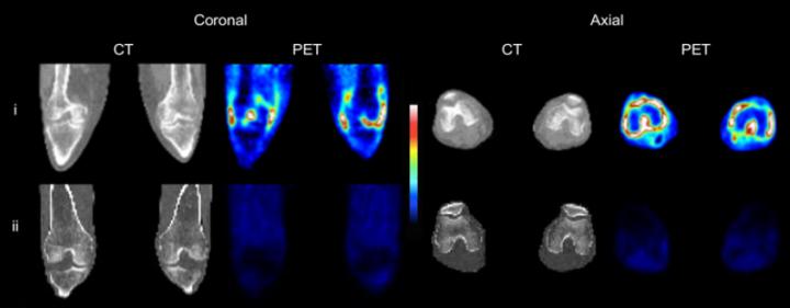
The study included three RA patients and three healthy volunteers who underwent PET scans of both knees using the TSPO radioligand carbon-11 (11C)-PBR28. In addition, cellular expression of TSPO was examined in synovial tissue from these individuals, plus three more RA patients and three more healthy patients (undergoing knee arthroscopy for injuries). TSPO mRNA expression and hydrogen-3 (3H)-PBR28 radioligand binding were assessed using in vitro monocytes, macrophages, fibroblast-like synoviocytes (FLS) and CD4+ T-lymphocytes.
Results showed that the 11C-PBR28 PET signal was significantly higher in RA joints compared to healthy joints. In addition, 3H-PBR28 specific binding in synovial tissue was approximately 10-fold higher in RA patients compared to healthy controls. Immunofluorescence revealed TSPO expression on macrophages, FLS and CD4+ T cells. In vitro study demonstrated highest TSPO mRNA expression and 3H-PBR28 specific binding in activated FLS, non-activated and activated 'M2' reparative macrophages. The lowest TSPO expression was in activated and non-activated CD4+ T lymphocytes.
Narayan notes, "It is well recognised that not all currently available treatments are capable of controlling joint inflammation in all patients with rheumatoid arthritis, hence the need to develop new pharmacological therapies. This work demonstrates that TSPO PET is able to act as a means of imaging not only synovial macrophages, but also activated synovial fibroblasts. The crucial role of the fibroblast and its soluble products in RA pathogenesis is increasingly realised."
She adds, "Indeed, there has been recent interest in targeting activated fibroblasts as a novel targeted treatment strategy for RA. Therefore, TSPO PET imaging in early phase clinical trials may provide a sensitive indication of treatment response to such novel therapies with a view to informing the design of later stage clinical trials. As our knowledge of cellular TSPO expression and behavior grows, TSPO-targeted imaging may also give us unique insights into the pathogenesis of inflammatory disease."
###
Authors of "Translocator protein as an imaging marker of macrophage and stromal activation in RA pannus" include Nehal Narayan, Harpreet Mandhair, Francesco Carlucci, Stephanie G. Dakin, Afsie Sabokbar, and Peter C. Taylor, University of Oxford, Headington, Oxford, UK; David R. Owen, Imperial College, Hammersmith, London, UK; Erica Smyth, Azeem Saleem, Roger N. Gunn, and Lisa Wells, Imanova Centre for Imaging Sciences, Hammersmith, London, UK; and Eugenii A. Rabiner, Imanova Centre for Imaging Sciences, Hammersmith, London, and King's College, London, UK.
The research was supported by the National Institute for Health Research Oxford Biomedical Research Centre, an IMPETUS pilot study grant from Imanova, Academic Centre for Imaging Sciences, an MRC clinician scientist award (MR/N008219/1), and Arthritis Research UK (20506).
Please visit the SNMMI Media Center to view the PDF of the study, including images, and more information about molecular imaging and personalized medicine. To schedule an interview with the researchers, please contact Laurie Callahan at (703) 652-6773 or [email protected]. Current and past issues of The Journal of Nuclear Medicine can be found online at http://jnm.snmjournals.org.
ABOUT THE SOCIETY OF NUCLEAR MEDICINE AND MOLECULAR IMAGING
The Society of Nuclear Medicine and Molecular Imaging (SNMMI) is an international scientific and medical organization dedicated to advancing nuclear medicine and molecular imaging, vital elements of precision medicine that allow diagnosis and treatment to be tailored to individual patients in order to achieve the best possible outcomes.
SNMMI's more than 15,000 members set the standard for molecular imaging and nuclear medicine practice by creating guidelines, sharing information through journals and meetings and leading advocacy on key issues that affect molecular imaging and therapy research and practice. For more information, visit http://www.snmmi.org.
Media Contact
Laurie F Callahan
[email protected]
@SNM_MI
http://www.snm.org
Original Source
http://www.snmmi.org/NewsPublications/NewsDetail.aspx?ItemNumber=29526 http://dx.doi.org/10.2967/jnumed.117.202200


