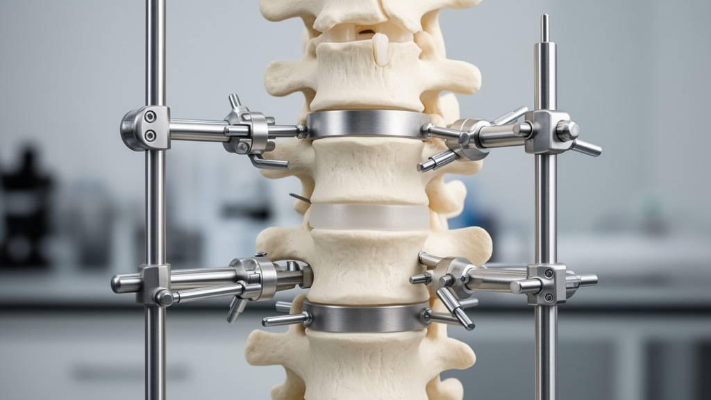
A computational biomechanics investigation has demonstrated that combining percutaneous pedicle screw fixation with a newly developed micro-adjustable external fixation system significantly alters spinal kinematics and reduces mechanical stress compared with standard internal screw fixation in simulated thoracolumbar burst fractures. Using three-dimensional finite element modeling of an L1 vertebral fracture, researchers analyzed how the experimental stabilization assembly stabilizes the injured spine under multiple loading directions. The simulation provides theoretical structural evidence assessing the device’s capability to limit postoperative loss of reduction and mitigate implant strain during minimally invasive spinal interventions.
To evaluate the structural performance of the configuration, the study authors developed two comparative computational cases representing an L1 burst fracture scenario. The first setup, designated as CASE 1, integrated the micro-adjustable external fixation framework, abbreviated as MAEF, alongside percutaneous pedicle screw stabilization. The second configuration, CASE 2, modeled percutaneous short-segment intermediate screw fixation, referred to as PSISF, representing a conventional surgical stabilization technique. The investigative protocol applied controlled loading conditions across both digital models to assess spinal mobility and load distribution across critical structural segments.
Biomechanical evaluation relied on two primary metrics measured under identical load-control protocols before and after instrumented fixation. The authors recorded the percentage change in range of motion across functional spinal segments and documented the maximum stress values sustained by the hardware assemblies and neighboring anatomical structures. Specific anatomical sites evaluated for stress concentrations included the fixation systems themselves, the fractured L1 vertebral body, the inferior endplate of the adjacent T12 vertebra, and the superior endplate of the adjacent L2 vertebra.
Analysis of range of motion revealed pronounced differences in structural stabilization between the two modeled interventions under all tested loading vectors. When normalized against an intact T11 to L3 anatomical segment model, the range of motion for the micro-adjustable external fixation framework was constrained to between 8.4 percent and 15.5 percent. In contrast, the percutaneous short-segment intermediate screw fixation model, normalized against an intact T12 to L2 reference model, preserved a substantially wider range of motion, recording values ranging from 48.0 percent to 70.9 percent across the applied load conditions.
Stress distribution assessments demonstrated that the micro-adjustable external fixation framework generally exhibited lower maximum stresses than the short-segment internal configuration across multiple evaluated structures. Lower peak stresses were recorded within the fixation hardware, the fractured L1 vertebra, the inferior endplate of T12, and the superior endplate of L2 under the computational loads. The highest stress levels in both fixation systems emerged during axial rotation maneuvers, with the external frame peaking at 77.025 megapascals during right lateral rotation, whereas the internal intermediate screw system reached a peak of 170.97 megapascals during left lateral rotation.
A notable divergence between the two stabilization models occurred in the stress profiles observed within the fractured L1 vertebral body itself. Within the micro-adjustable external fixation model, the fractured vertebra sustained markedly higher maximum stress levels during flexion and extension cycles than during lateral bending or axial rotational movements. These vertebral body stresses under flexion and extension reached magnitudes comparable to those registered in the percutaneous short-segment model, a biomechanical response that the authors attributed directly to the mechanical load-sharing characteristics of the external frame’s micro-adjustable mechanism.
Mechanical demands placed on neighboring uninjured anatomy also differed markedly between the tested computational systems. Under every simulated loading condition, the maximum stress concentrations observed across the adjacent vertebral endplates—specifically the T12 inferior and L2 superior endplates—remained consistently lower in the micro-adjustable external fixation model than in the short-segment screw configuration.
The study authors concluded that the biomechanical features demonstrated by the micro-adjustable external frame offer theoretical structural support for its potential use in minimally invasive thoracolumbar burst fracture management. By providing robust segmental rigidity while reducing peak stress concentrations on the fixation hardware and adjacent endplates, the system’s mechanical profile may theoretically reduce risks of implant failure, decrease post-traumatic kyphotic deformity progression, and facilitate spinal fracture healing, though clinical translation remains dependent on prospective empirical validation.
Subject of Research: Biomechanical finite element analysis of a micro-adjustable external fixation frame for thoracolumbar burst fractures
Article Title: Biomechanical study of a novel external fixation frame controlling postoperative loss of reduction in thoracolumbar fractures: Finite element analysis
Article References: Guo, H., Feng, J., Xue, Y., Wang, K., Hao, M., & Nie, S. (2026). Biomechanical study of a novel external fixation frame controlling postoperative loss of reduction in thoracolumbar fractures: Finite element analysis. BMC Musculoskeletal Disorders. https://doi.org/10.1186/s12891-026-10720-4
Image Credits: AI Generated
DOI: 10.1186/s12891-026-10720-4
Keywords: finite element analysis, thoracolumbar fractures, external fixation, biomechanics, pedicle screw fixation, orthopedic surgery, spinal trauma, burst fractures, vertebral fracture, range of motion, stress distribution, lumbar spine, thoracic spine, implant failure, kyphosis, orthopedic devices
News Source: Ophelia Keating. (November 12, 2026). Novel External Fixation Frame Reduces Spinal Stress in Finite Element Analysis. Scienmag.