• HOME
  • NEWS
  • EXPLORE
    • CAREER
      • Companies
      • Jobs
    • EVENTS
    • iGEM
      • News
      • Team
    • PHOTOS
    • VIDEO
    • WIKI
  • BLOG
  • COMMUNITY
    • FACEBOOK
    • INSTAGRAM
    • TWITTER
Friday, August 1, 2025
BIOENGINEER.ORG
No Result
View All Result
  • Login
  • HOME
  • NEWS
  • EXPLORE
    • CAREER
      • Companies
      • Jobs
        • Lecturer
        • PhD Studentship
        • Postdoc
        • Research Assistant
    • EVENTS
    • iGEM
      • News
      • Team
    • PHOTOS
    • VIDEO
    • WIKI
  • BLOG
  • COMMUNITY
    • FACEBOOK
    • INSTAGRAM
    • TWITTER
No Result
View All Result
Bioengineer.org
No Result
View All Result
Home NEWS Science News Chemistry

Noninvasive, label-free optical method visualizes deep, cellular brain disease in vivo

Bioengineer by Bioengineer
July 16, 2021
in Chemistry
Reading Time: 2 mins read
0
Share on FacebookShare on TwitterShare on LinkedinShare on RedditShare on Telegram

IMAGE

Credit: by Jun Zhu, Hercules Rezende Freitas, Izumi Maezawa, Lee-way Jin, and Vivek J. Srinivasan

Central nervous system (CNS) diseases such as Alzheimer’s disease (AD) manifest early at the microscopic (i.e. cellular) level, deep in the brain. Yet, optical microscopes that can see cells in the living brain are superficial or invasive. Whole brain imaging techniques such as magnetic resonance imaging are deep and non-invasive, but lack cellular resolution.

In a new paper published in Light Science & Application, a team of scientists, led by Professor Vivek J. Srinivasan from the Departments of Ophthalmology and Radiology and Tech4Health Institute, NYU Langone Health, USA, and co-workers have developed a label-free optical microscopy approach that has a unique ability to image deep, with high resolution and minimal invasiveness. Specifically, they demonstrated an in vivo high numerical aperture optical coherence microscopy (OCM) approach that utilizes the 1700 nm water absorption window, where attenuation of light by scattering and absorption is minimized.

The 1700 nm water absorption window, also known as the third near-infrared (NIR) window, boasts a local water absorption minimum and relatively low scattering. In OCM, a broader spectrum provides a finer axial resolution, and with it, a stronger ability to reject multiply scattered light that causes image blur. Yet the entire 1700 nm window, which spans from 1560 to 1820 nm, is often not used:

“The transition from standard wavelengths to 1700 nm OCM, while optimally using the entire water absorption window (not just a portion of the window), has been very difficult to date due to the numerous optical engineering challenges.” the scientists mentioned.

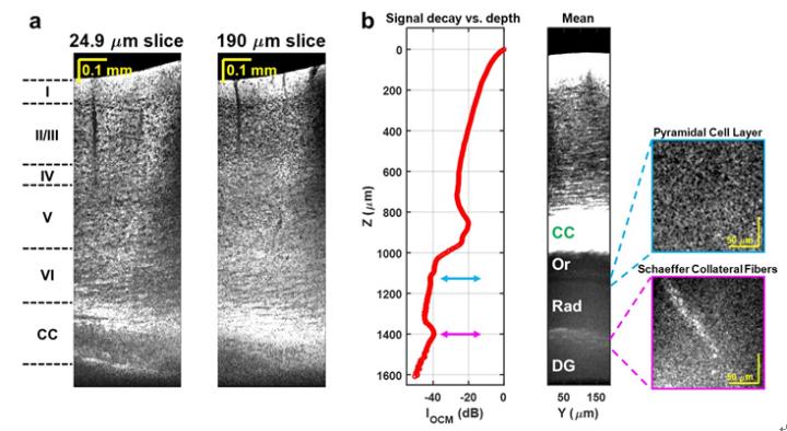
These challenges include noisy detectors and light sources, severe chromatic dispersion, and lack of standardized optical components. The scientists addressed these issues through the choice of a low noise supercontinuum light source, a custom numerical dispersion compensation method, and optical system design. With these technical advances, neuronal cell and myelin architecture across the entire depth of the mouse neocortex, and some sub-cortical regions, can be imaged through a thinned-skull preparation that preserves intracranial space.

“The results represent unprecedented depths for cellular-scale brain imaging through a minimally invasive preparation. We next investigated the 5xFAD mouse model of Alzheimer’s disease (AD), which is expected to show a gradation of pathology with cortical depth. The imaging results confirmed the appearance of severe pathology in deep but not superficial cortex, which would be missed by more superficial imaging techniques.”

Another important feature of the method is that the image contrast arises from intrinsic properties of the brain itself. OCM does not require transgenic mice or administration of compounds. Neuronal cell body loss, demyelination of axons, plaques, and local tissue changes can all be imaged.

“Now disease can be visualized deep in the mouse brain with a simple surgical preparation, without exogenous labeling. The 1700 nm optical window can also quantify tissue water and lipid content in vivo, which may provide further insights into disease progression.” the scientists forecast.

###

Media Contact
Jun Zhu
[email protected]

Related Journal Article

http://dx.doi.org/10.1038/s41377-021-00586-7

Tags: Chemistry/Physics/Materials SciencesOptics
Share13Tweet8Share2ShareShareShare2

Related Posts

blank

Nanodevice Harnesses Sound Waves to Shape Light, Revolutionizing Displays and Imaging Technologies

August 1, 2025
Here’s a rewritten version of the headline for a science magazine post: “Could Desert Dust Hold the Key to Freezing Clouds?”

Here’s a rewritten version of the headline for a science magazine post: “Could Desert Dust Hold the Key to Freezing Clouds?”

July 31, 2025

Rice Theoretical Physicist Illuminates Rare High-Field Phase in Superconductivity Research

July 31, 2025

Sunlight Transforms the Chemical Breakdown of Discarded Face Masks

July 31, 2025
Please login to join discussion

POPULAR NEWS

  • Blind to the Burn

    Overlooked Dangers: Debunking Common Myths About Skin Cancer Risk in the U.S.

    60 shares
    Share 24 Tweet 15
  • Dr. Miriam Merad Honored with French Knighthood for Groundbreaking Contributions to Science and Medicine

    46 shares
    Share 18 Tweet 12
  • Study Reveals Beta-HPV Directly Causes Skin Cancer in Immunocompromised Individuals

    37 shares
    Share 15 Tweet 9
  • Engineered Cellular Communication Enhances CAR-T Therapy Effectiveness Against Glioblastoma

    35 shares
    Share 14 Tweet 9

About

We bring you the latest biotechnology news from best research centers and universities around the world. Check our website.

Follow us

Recent News

Developing Innovative Flexible Materials for Self-Powered Wearable Sensors

Cutting-Edge Wound Monitor Set to Revolutionize Chronic Infection Management

Can Specific Fungi Enhance the Micronutrient Levels in Bread Wheat?

  • Contact Us

Bioengineer.org © Copyright 2023 All Rights Reserved.

Welcome Back!

Login to your account below

Forgotten Password?

Retrieve your password

Please enter your username or email address to reset your password.

Log In
No Result
View All Result
  • Homepages
    • Home Page 1
    • Home Page 2
  • News
  • National
  • Business
  • Health
  • Lifestyle
  • Science

Bioengineer.org © Copyright 2023 All Rights Reserved.