Reston, VA—The protein galectin-1 (Gal-1) has been identified as a new PET imaging biomarker for immune checkpoint blockade (ICB) therapy, allowing physicians to predict the tumor responses before beginning treatment. Information garnered from Gal-1 PET imaging could also be used to facilitate patient stratification and optimize immunotherapy, enabling targeted interventions and improving patient outcomes. This research was published in the May issue of The Journal of Nuclear Medicine.

Credit: Image created by N Liu and X Yang, et al. Peking University, Beijing, China.
Reston, VA—The protein galectin-1 (Gal-1) has been identified as a new PET imaging biomarker for immune checkpoint blockade (ICB) therapy, allowing physicians to predict the tumor responses before beginning treatment. Information garnered from Gal-1 PET imaging could also be used to facilitate patient stratification and optimize immunotherapy, enabling targeted interventions and improving patient outcomes. This research was published in the May issue of The Journal of Nuclear Medicine.
Immunotherapies, such as ICB, have produced promising clinical outcomes in melanoma, non–small cell lung cancer, and several other types of tumors. However, only a subgroup of patients experiences positive outcomes with objective response rates spanning between five and 60 percent.
“Developing reliable approaches for assessing responses and selecting eligible patients for immunotherapy remains challenging,” said Zhaofei Liu, PhD, Boya Distinguished Professor at Peking University in Beijing, China. “Current clinical criteria for monitoring solid tumor responses to immunotherapy are based on CT and MRI scans, but these methods result in a considerable delay between treatment commencement and response evaluation. Molecular imaging techniques, especially PET, have emerged as robust tools for predicting immunotherapy effectiveness through the real-time, quantitative, and noninvasive assessment of biomarkers in vivo.”
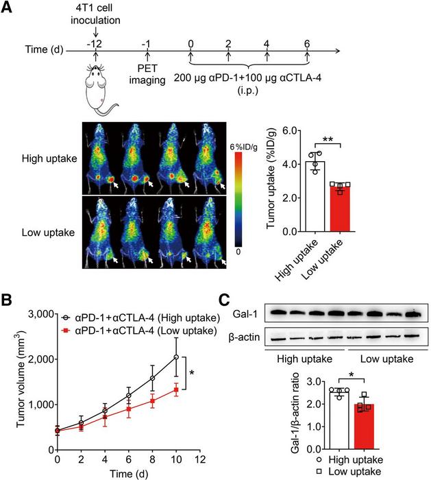
In the study, a mouse model was utilized to identify new imaging biomarkers for tumor responses to ICB therapy. Through a proteomic analysis (separation, identification, and quantification of proteins in a tumor), researchers found that tumors exhibiting low Gal-1 expression responded positively to ICB therapy.
Next, Gal-1 was labeled with 124I and the radiotracer (124I-α-Gal-1) and small animal PET imaging and biodistribution studies were conducted to assess the specificity of the radiotracer. PET imaging with 124I-αGal-1 showed the immunosuppressive status of the tumor microenvironment, thus enabling the prediction of ICB resistance in advance of treatment. For tumors that were not predicted to respond well to ICB therapy, researchers developed a rescue strategy that utilized a Gal-1 inhibitor that significantly improved the chance for success.
“Gal-1 PET opens avenues for the early prediction of ICB efficacy before treatment initiation and facilitates the precision design of combinational regimes,” noted Liu. “This sensitive approach has the potential to achieve individualized precision treatment for patients in the future.”
This research was published online in March 2024.
The authors of “Noninvasively Deciphering the Immunosuppressive Tumor Microenvironment Using Galectin-1 PET to Inform Immunotherapy Responses” include Ning Liu, Xiujie Yang, Chao Gao, Jianze Wang, Yuwen Zeng, Linyu Zhang, Qi Yin, Ting Zhang, Haoyi Zhou, Kui Li, and Jinhong Du, Department of Radiation Medicine, School of Basic Medical Sciences, Peking University Health Science Center, Beijing, China; Shixin Zhou, Department of Cell Biology, School of Basic Medical Sciences, Peking University Health Science Center, Beijing, China; Xuyang Zhao, Institute of Systems Biomedicine, Department of Pathology, School of Basic Medical Sciences, Peking University Health Science Center, Beijing, China; Hua Zhu and Zhi Yang, Key Laboratory of Carcinogenesis and Translational Research (Ministry of Education/Beijing), Department of Nuclear Medicine, Peking University Cancer Hospital and Institute, Beijing, China; and Zhaofei Liu, Key Laboratory of Carcinogenesis and Translational Research (Ministry of Education/Beijing), Department of Nuclear Medicine, Peking University Cancer Hospital and Institute, Beijing, China, Department of Nuclear Medicine, Peking University Third Hospital, Beijing, China, State Key Laboratory of Vascular Homeostasis and Remodeling, Peking University, Beijing, China, and Peking University–Yunnan Baiyao International Medical Research Center, Beijing, China.
Visit the JNM website for the latest research, and follow our new Twitter and Facebook pages @JournalofNucMed or follow us on LinkedIn.
###
Please visit the SNMMI Media Center for more information about molecular imaging and precision imaging. To schedule an interview with the researchers, please contact Rebecca Maxey at (703) 652-6772 or [email protected].
About JNM and the Society of Nuclear Medicine and Molecular Imaging
The Journal of Nuclear Medicine (JNM) is the world’s leading nuclear medicine, molecular imaging and theranostics journal, accessed more than 16 million times each year by practitioners around the globe, providing them with the information they need to advance this rapidly expanding field. Current and past issues of The Journal of Nuclear Medicine can be found online at http://jnm.snmjournals.org.
JNM is published by the Society of Nuclear Medicine and Molecular Imaging (SNMMI), an international scientific and medical organization dedicated to advancing nuclear medicine and molecular imaging—precision medicine that allows diagnosis and treatment to be tailored to individual patients in order to achieve the best possible outcomes. For more information, visit www.snmmi.org.
Journal
Journal of Nuclear Medicine
DOI
10.2967/jnumed.123.266888
Article Title
Noninvasively Deciphering the Immunosuppressive Tumor Microenvironment Using Galectin-1 PET to Inform Immunotherapy Responses
Article Publication Date
1-May-2024
COI Statement
This work was supported by the National Key R&D Program of China (2023YFC3404600 to Zhaofei Liu), the National Natural Science Foundation of China (81920108020 and 82325028 to Zhaofei Liu), the Beijing Natural Science Foundation (Z220011 and Z220014 to Zhaofei Liu), and the Beijing Nova Program Interdisciplinary Cooperation Project (20220484182 to Hua Zhu and Zhaofei Liu).


