• HOME
  • NEWS
  • EXPLORE
    • CAREER
      • Companies
      • Jobs
    • EVENTS
    • iGEM
      • News
      • Team
    • PHOTOS
    • VIDEO
    • WIKI
  • BLOG
  • COMMUNITY
    • FACEBOOK
    • INSTAGRAM
    • TWITTER
Friday, September 26, 2025
BIOENGINEER.ORG
No Result
View All Result
  • Login
  • HOME
  • NEWS
  • EXPLORE
    • CAREER
      • Companies
      • Jobs
        • Lecturer
        • PhD Studentship
        • Postdoc
        • Research Assistant
    • EVENTS
    • iGEM
      • News
      • Team
    • PHOTOS
    • VIDEO
    • WIKI
  • BLOG
  • COMMUNITY
    • FACEBOOK
    • INSTAGRAM
    • TWITTER
No Result
View All Result
Bioengineer.org
No Result
View All Result
Home NEWS Science News Biology

Newly discovered infectious prion structure shines light on mad cow disease

Bioengineer by Bioengineer
January 25, 2018
in Biology, Science News
Reading Time: 2 mins read
0
Share on FacebookShare on TwitterShare on LinkedinShare on RedditShare on Telegram
IMAGE

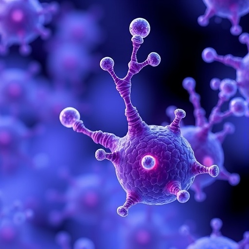
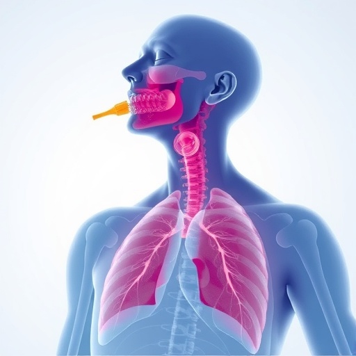
(Edmonton, AB) Groundbreaking research from the University of Alberta has identified the structure of the infectious prion protein, the cause of "mad cow disease" or BSE, chronic wasting disease in deer and elk and Creutzfeldt-Jakob disease in humans, which has long remained a mystery.

The infectious prion protein is a misfolded protein, which makes it very difficult to purify and study. Since it clumps together, standard structural biology techniques cannot be used to study it. Since the protein was first purified in the 1980s researchers have made limited insights into the structure of the protein.

The collaborative study, published in PLOS Pathogens, used electron cryomicroscopy to collect high-resolution electron micrographs. This was the first time this technology has been used on amyloid fibrils of the infectious prion, which are a special form of clumped-together proteins that form fibrils.

"The recent advances to electron cryomicroscopy technology are certainly a breakthrough," says Holger Wille, co-principal investigator and an associate professor in the Department of Biochemistry at the University of Alberta's Faculty of Medicine & Dentistry. "We know the structure of the healthy normal cellular form of the protein, but we knew very little about the infectious prion protein and how it propagates. The use of these high-powered microscopes has finally given us some clarity."

The team had to develop a processing scheme for the data masses. There were thousands of electron micrographs, and they had to extract the best images. After three years of working, developing techniques and processing data the results in the paper are a three-dimensional model for the structure of the infectious prion protein.

"It is not an atomistic model, so we cannot say which position the atoms are in," says Wille. "But this is something we hope to do in the future."

The model can give insights into how the infectious prion protein propagates. The structure argues against existing theories of prion conversion and suggests how the process might actually work. The study suggests how infectious prions replicate by converting non-infectious, cellular versions into copies of themselves.

Moving forward, the researchers want to go into more depth. This study used model system prions, but they are now using the prions that infect cows (BSE), wild animals (Chronic Wasting Disease) and humans (Creutzfeldt-Jakob Disease).

"Ultimately, if we know how the prion propagates, we could come up with clinical interventions to treat or prevent disease," says Wille.

###

This research was supported by the Alberta Prion Research Institute and the Alberta Livestock and Meat Agency.

"The Structural Architecture of an Infectious Mammalian Prion Using Electron Cryomicroscopy," PLOS Pathogens, published online September 8, 2016.

Media Contact

Shelby Soke
[email protected]
403-988-4730
@ualberta_fomd

http://www.med.ualberta.ca

Share12Tweet7Share2ShareShareShare1

Related Posts

blank

Coherent Detector Measures Vectorial Light Non-Separability

September 26, 2025

Hormones Shape Brain Structure Across Menstrual Cycle

September 26, 2025

Classifying Malignant Colon Polyps via OCT

September 26, 2025

Caspase-8–Meteorin Roles in MASH, Fibrosis

September 26, 2025
Please login to join discussion

POPULAR NEWS

  • New Study Reveals the Science Behind Exercise and Weight Loss

    New Study Reveals the Science Behind Exercise and Weight Loss

    79 shares
    Share 32 Tweet 20
  • Physicists Develop Visible Time Crystal for the First Time

    72 shares
    Share 29 Tweet 18
  • Scientists Discover and Synthesize Active Compound in Magic Mushrooms Again

    55 shares
    Share 22 Tweet 14
  • Tailored Gene-Editing Technology Emerges as a Promising Treatment for Fatal Pediatric Diseases

    51 shares
    Share 20 Tweet 13

About

We bring you the latest biotechnology news from best research centers and universities around the world. Check our website.

Follow us

Recent News

Coherent Detector Measures Vectorial Light Non-Separability

Hormones Shape Brain Structure Across Menstrual Cycle

Classifying Malignant Colon Polyps via OCT

  • Contact Us

Bioengineer.org © Copyright 2023 All Rights Reserved.

Welcome Back!

Login to your account below

Forgotten Password?

Retrieve your password

Please enter your username or email address to reset your password.

Log In
No Result
View All Result
  • Homepages
    • Home Page 1
    • Home Page 2
  • News
  • National
  • Business
  • Health
  • Lifestyle
  • Science

Bioengineer.org © Copyright 2023 All Rights Reserved.