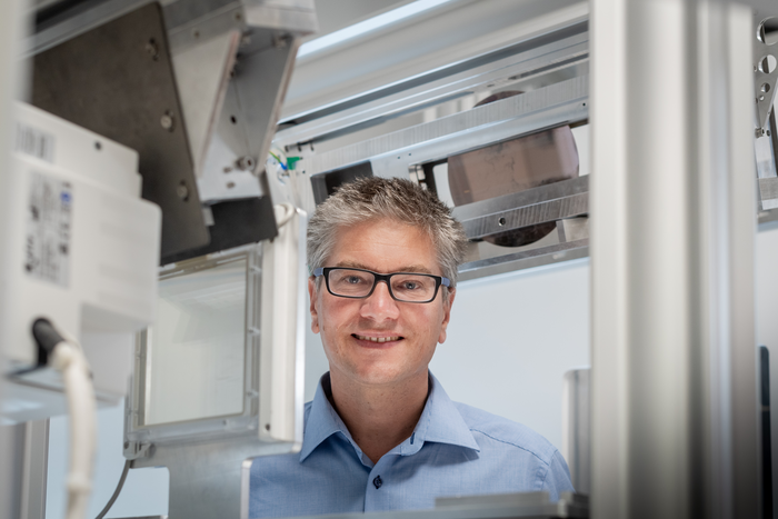
A research team at the Technical University of Munich (TUM) has, for the first time, produced dark-field X-ray images of patients infected with the corona virus. In contrast to conventional X-ray images, dark-field images visualize the microstructure of the lung tissue, thereby providing additional information. This approach has the potential to provide an alternative to computed tomography (CT), which requires a significantly higher radiation dose.
Credit: Andreas Heddergott / TUM
A research team at the Technical University of Munich (TUM) has, for the first time, produced dark-field X-ray images of patients infected with the corona virus. In contrast to conventional X-ray images, dark-field images visualize the microstructure of the lung tissue, thereby providing additional information. This approach has the potential to provide an alternative to computed tomography (CT), which requires a significantly higher radiation dose.
The lungs of Covid-19 patients are normally visualized using computed tomography (CT). CT technology uses multiple X-ray images from different angles to compute a three-dimensional image. This provides more accurate results than two-dimensional imaging using conventional X-ray technology. The downside, however, is a higher radiation dose due to the large number of X-ray images required.
Dark-field chest X-ray is a new X-ray technology developed by Prof. Franz Pfeiffer. It is paving the road for new possibilities in radiological diagnostics: “During our X-ray examination, we take conventional X-ray images and dark-field images simultaneously. This gives us additional information about the affected lung tissue quickly and easily,” says Franz Pfeiffer, Professor of Biomedical Physics and Director of the Munich Institute of Biomedical Engineering at TUM.
“The resulting radiation dose is fifty times smaller compared to CT machines. Consequently, this method is promising for application scenarios that require repeated examinations over extended periods of time – for example, when investigating the progression of long covid. This approach might serve as an alternative to computed tomography for imaging lung tissue over prolonged observation periods,” Franz Pfeiffer explains further.
Additional information on lung tissue microstructure
In a new study, radiologists compared the images of patients with Covid 19 lung disease to those of healthy individuals. They found that distinguishing between sick and healthy individuals was easier using dark-field images than conventional X-ray images. The radiologists were able to differentiate between diseased and healthy lung tissue most readily when both kinds of images – conventional and darkfield – were available.
While conventional X-ray technology relies on the attenuation of X-rays, dark-field X-ray technology utilizes so-called small-angle X-ray scattering. This opens the door to garner additional information on the nature of lung tissue microstructure. Dark-field images can thus provide added value when investigating a variety of lung diseases.
Quantitative evaluation
The research team optimized the X-ray machine prototype to allow them to evaluate the images quantitatively. A healthy lung with many intact alveoli produces a strong dark-field signal and appears bright in the image. In contrast, inflamed lung tissue, with embedded fluid, produces a weaker signal and appears darker in the image. “We normalize the dark-field signal with regard to lung volume to account for lung volume variations between individuals,” explains Manuela Frank, a lead author of the paper.
“Next, we hope to examine further patients. Once we have sufficient dark-field X-ray data available, we intend to use artificial intelligence to support the evaluation process. For conventional images, we have already carried out a pilot project for AI evaluation of our X-ray images,” says Daniela Pfeiffer, professor of radiology and medical director of the study at the TUM University hospital Klinikum rechts der Isar.
More information on the new dark-field X-ray technology:
Dark-field X-ray is a novel medical examination method. Prof. Franz Pfeiffer and his team developed the method from the ground up and have been advancing it continuously for over ten years, making it available for the world-wide first use in patients.
In close cooperation with radiologists at the TUM University hospital Klinikum rechts der Isar, the researchers developed the dark-field X-ray prototype which has been approved for initial clinical studies. The prototype is currently being used in several studies with patients suffering from various lung diseases. Following an initial study on chronic obstructive pulmonary disease (COPD) and Covid-19, the technology might also be deployed in the future for other lung pathologies such as lung cancer, fibrosis, or pneumothorax.
- Grating based x-ray dark field imaging – How the method works
- Schematic representation (illustration)
- New technology for clinical CT scans (2022)
- New X-ray technology first used with patients (2021) – First use with patients with the lung disease COPD
Further information:
Prof. Franz Pfeiffer is the Director of the Munich Institute of Biomedical Engineering (MIBE). MIBE is an Integrative Research Institute (IRI) within the Technical University of Munich (TUM). At MIBE, researchers specializing in medicine, the natural sciences, and engineering join forces to develop new methods for diagnosing or treating diseases. They also work on improving technologies that compensate for physical disabilities. The activities cover the entire development process – from the study of basic scientific principles through to their application in new medical devices, medicines and software.
The authors Franz Pfeiffer (TUM), Daniela Pfeiffer (TUM) and Thomas Koehler (Philips Research) are Fellows of the TUM Institute for Advanced Study (TUM-IAS). The TUM-IAS creates interdisciplinary research groups with researchers from TUM and guests from other research institutions and industry to explore new, challenging fields of research.
The work was supported by the European Research Council through a Starting and an Advanced Grant, and by Philips by providing technical components.
Journal
Communications Medicine
DOI
10.1038/s43856-022-00215-3
Method of Research
Randomized controlled/clinical trial
Subject of Research
People
Article Title
Dark-field chest X-ray imaging for the assessment of COVID-19-pneumonia
Article Publication Date
21-Nov-2022
COI Statement
T.K. and K.J.E. are employees of Philips GmbH Innovative Technologies. The remaining authors declare no competing interests.
