• HOME
  • NEWS
  • EXPLORE
    • CAREER
      • Companies
      • Jobs
    • EVENTS
    • iGEM
      • News
      • Team
    • PHOTOS
    • VIDEO
    • WIKI
  • BLOG
  • COMMUNITY
    • FACEBOOK
    • INSTAGRAM
    • TWITTER
Wednesday, September 10, 2025
BIOENGINEER.ORG
No Result
View All Result
  • Login
  • HOME
  • NEWS
  • EXPLORE
    • CAREER
      • Companies
      • Jobs
        • Lecturer
        • PhD Studentship
        • Postdoc
        • Research Assistant
    • EVENTS
    • iGEM
      • News
      • Team
    • PHOTOS
    • VIDEO
    • WIKI
  • BLOG
  • COMMUNITY
    • FACEBOOK
    • INSTAGRAM
    • TWITTER
No Result
View All Result
Bioengineer.org
No Result
View All Result
Home NEWS Science News Chemistry

New way of optical visualization of nano objects proposed

Bioengineer by Bioengineer
March 29, 2019
in Chemistry
Reading Time: 2 mins read
0
IMAGE
Share on FacebookShare on TwitterShare on LinkedinShare on RedditShare on Telegram

A paper appeared in Nanoscale in March 2019

IMAGE

Credit: Kazan Federal University

High-resolution optical microscopy methods promise breakthroughs in materials science, biology, and medicine. Today, their possibilities basically reach those of scanning electron microscopy.

Sergey Kharintsev, Head of Nano-Optics Laboratory (Kazan Federal University), comments, “The main advantage of optical microscopy is that it provides nondestructive chemical analysis of single molecules exposed to continuous-wave low-powered laser light. Unlike scanning electron microscopy, this allows one to perform 3D visualization and spectral analysis of intrinsic structure of nano objects, and also get insights into processes in living cells.”

According to Professor Kharintsev, fluorescence microscopy has become the most popular method for biology and medicine lately. Its main drawback is the need for fluorescent labels that must be photostable and non-toxic. Furthermore, there are factors of labor-intensive sample preparation and fluorophore prices.

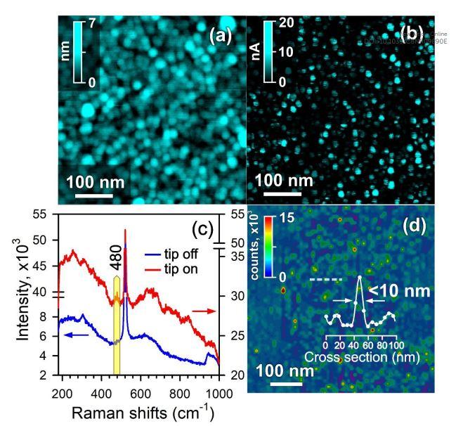
The paper posits that one of the solutions for ultrahigh resolution microscopy is a superlens, first proposed by John Pendry (Imperial College London, UK) in 2000. The lens looks like a sandwich with a metallic film placed between two dielectric layers. Superresolution is achieved due to the optical near-field enhancement through surface plasmon resonances. Lately, a breakthrough initiative in practical implementation of such superlens has been put forth by Vladimir Shalaev (Purdue University, USA), who suggested using a nano-composite metal-dielectric film. This permits the superlens to be operated at a tunable single frequency.

“We propose to use a nano-structured metal-dielectric film that exhibits a double epsilon-near-zero behavior near the percolation threshold,” continues Sergey Kharintsev. “The material in question is titanium oxynitride, a compound first synthesized by Andrei Mihai’s group at Imperial College London. Such a superlens provides ultrahigh spatial resolution due to stimulated Raman scattering. Consequently, we have succeeded to achieve a spatial resolution of 8 nm and 80 nm in the near-field and far-field, respectively. Importantly, obtained optical images are formed with a standard objective only, not using optical nano-antennas, designed laser beams, or fluorescent labels.”

###

The research has been supported by the Russian Science Foundation. The project’s title is “Synthesis and research of ultra-thin magnetic heterostructures with potential spintronic and optronic applications”; Professor Lenar Tagirov (Institute of Physics, Kazan Federal University) is at the helm. Sergey Kharintsev’s PhD student Anton Kharitonov has prepared his PhD thesis based on the results.

Media Contact
Yury Nurmeev
[email protected]

Original Source

https://kpfu.ru/eng/news-eng/new-way-of-optical-visualization-of-nano-objects.html

Related Journal Article

http://dx.doi.org/10.1039/C8NR09890E

Tags: Chemistry/Physics/Materials SciencesOptics
Share24Tweet7Share2ShareShareShare1

Related Posts

Oxford AI Tool Revolutionizes Supernova Discovery Amidst Cosmic Noise

Oxford AI Tool Revolutionizes Supernova Discovery Amidst Cosmic Noise

September 10, 2025
Innovative Methods for Generating Methanol Using Electricity and Biomass

Innovative Methods for Generating Methanol Using Electricity and Biomass

September 9, 2025

Isotope Tafel Analysis Reveals Proton Transfer Kinetics

September 9, 2025

Gemini South Uncovers Elusive Cloud-Forming Chemical on Ancient Brown Dwarf

September 9, 2025
Please login to join discussion

POPULAR NEWS

  • blank

    Breakthrough in Computer Hardware Advances Solves Complex Optimization Challenges

    151 shares
    Share 60 Tweet 38
  • New Drug Formulation Transforms Intravenous Treatments into Rapid Injections

    116 shares
    Share 46 Tweet 29
  • Physicists Develop Visible Time Crystal for the First Time

    52 shares
    Share 21 Tweet 13
  • First Confirmed Human Mpox Clade Ib Case China

    56 shares
    Share 22 Tweet 14

About

We bring you the latest biotechnology news from best research centers and universities around the world. Check our website.

Follow us

Recent News

Scientists Discover Giant DNA Hidden Within the Human Mouth

Fermented Poncirus Extract Inhibits Fat Cell Formation

Breakthrough: First-Ever Koala Chlamydia Vaccine Receives Approval

  • Contact Us

Bioengineer.org © Copyright 2023 All Rights Reserved.

Welcome Back!

Login to your account below

Forgotten Password?

Retrieve your password

Please enter your username or email address to reset your password.

Log In
No Result
View All Result
  • Homepages
    • Home Page 1
    • Home Page 2
  • News
  • National
  • Business
  • Health
  • Lifestyle
  • Science

Bioengineer.org © Copyright 2023 All Rights Reserved.