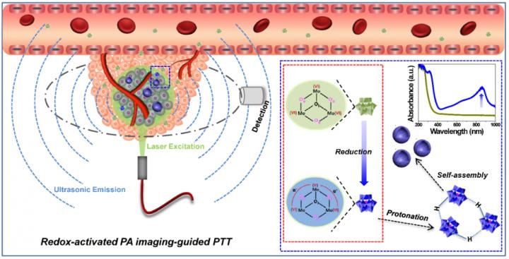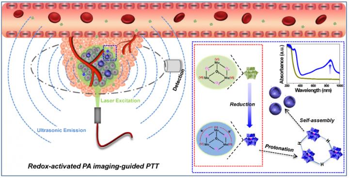
Credit: D. Ni and W. Cai et al., Department of Radiology, University of Wisconsin-Madison, Madison, Wisc.
PHILADELPHIA – A novel, intelligent theranostic agent for precise tumor diagnosis and therapy has been developed that remains as small molecules while circulating in the bloodstream, can then self-assemble into larger nanostructures in the tumor, and be activated by the tumor microenvironment for therapy guided by photoacoustic imaging. The research was presented at the 2018 Annual Meeting of the Society of Nuclear Medicine and Molecular Imaging (SNMMI).
"Although various types of imaging agents have been developed for photoacoustic (PA) imaging, relatively few imaging agents exhibit high selectivity to the tumor microenvironment for on-demand PA imaging and therapy," Dalong Ni, PhD, a postdoctoral scholar in the Molecular Imaging and Nanotechnology Laboratory at the University of Wisconsin-Madison (Website: http://mi.wisc.edu; PI: Weibo Cai, PhD), explains. "In this study, an intelligent theranostic agent of molybdenum-based polyoxometalate clusters (denoted as Ox-POM) was designed. These clusters work like an intelligent "nano-robot" in vivo, first searching the tumor area (which can be noninvasively images with PA imaging) and then killing tumor cells (photothermal therapy after it self-assembled in the tumor microenvironment)."
Ni points out, "Unlike traditional chemotherapy, the designed intelligent Ox-POM clusters, obtained from an easy, fast, and large-scale synthesis process, will only cause damage in tumor areas but not to the normal tissues and organs. Importantly, like most clinical imaging agents, these nanoclusters are mainly excreted through the kidneys, making them highly biocompatible and reducing the potential toxic effects on patients."
Tumor-bearing mice were tested with this novel system of redox-activated PA imaging-guided photothermal therapy (PTT). Redox is short for reduction-oxidation reaction. In this study, the ultra-small Ox-POM clusters accumulate in the tumor and are reduced in the tumor microenvironment. They then get protonated and self-assemble into much larger nanoparticles that are near-infrared (NIR) absorptive. Systematic in vitro and in vivo experiments were performed to evaluate their bioresponsive and theranostic capability.
Results from positron emission tomography (PET) imaging with zirconium-89-labeled Ox-POM showed these clusters could escape from recognition by the liver and spleen and were mainly excreted through the kidneys, which is highly desirable for reducing potential toxic effects. Studies in the tumor-bearing mice showed that the PA signal was detected in the tumors as early as one hour post-injection. Under laser irradiation, the temperature of the tumor rapidly increased, reaching above 40 °C in 30 seconds and reaching 52 °C in five minutes; the tumor growth was eliminated without subsequent recurrence for a prolonged period of up to 2 months, whereas the control groups demonstrated rapid tumor growth.
Ni states, "As a proof-of-concept, our findings explore a new strategy for precise tumor diagnosis and therapy, which is also expected to establish a new class of theranostic agents based on clusters, bridging the conventional concepts of "molecule" and "nano" in the bioimaging field." He adds, "These are exciting smart nanomaterials (target and/or respond to cancer and efficiently clear from the body) for potential clinical translation. On-demand tumor diagnosis and therapy triggered by physiological microenvironment characteristics of tumors can simultaneously reduce the damage of anticancer agents to normal organs/tissues and improve therapeutic efficacy."
###
Abstract 188: "Redox-Activated Photoacoustic Imaging-Guided Photothermal Therapy with Bioresponsive Polyoxometalate Cluster," Dalong NI, PhD, Dawei Jiang, PhD, Emily B. Ehlerding, Todd E. Barnhart, PhD, and Weibo Cai, PhD, University of Wisconsin – Madison, Madison, WI; Bo Yu, PhD, University of Wisconsin – Madison, Madison, WI, and School of Chemical Engineering and Pharmacy, Wuhan Institute of Technology, Wuhan, China; and Weijun Wei, Shanghai Jiao Tong University Affiliated Sixth People's Hospital, Shanghai, China. SNMMI 2018 Annual Meeting, June 23-26, 2018, Philadelphia.
Link to Abstract
Please visit the SNMMI Media Center for more information about molecular imaging and personalized medicine. To schedule an interview with the researchers, please contact Laurie Callahan at 703-652-6773 or [email protected]. 2018 SNMMI Annual Meeting abstracts can be found online at http://jnm.snmjournals.org/content/59/supplement_1. Current and past issues of The Journal of Nuclear Medicine are online at http://jnm.snmjournals.org.
ABOUT THE SOCIETY OF NUCLEAR MEDICINE AND MOLECULAR IMAGING
The Society of Nuclear Medicine and Molecular Imaging (SNMMI) is an international scientific and medical organization dedicated to advancing nuclear medicine and molecular imaging, vital elements of precision medicine that allow diagnosis and treatment to be tailored to individual patients in order to achieve the best possible outcomes.
SNMMI's more than 16,000 members set the standard for molecular imaging and nuclear medicine practice by creating guidelines, sharing information through journals and meetings and leading advocacy on key issues that affect molecular imaging and therapy research and practice. For more information, visit http://www.snmmi.org.
Media Contact
Laurie F Callahan
[email protected]
@SNM_MI
http://www.snm.org
Original Source
http://www.snmmi.org/NewsPublications/NewsDetail.aspx?ItemNumber=29469


