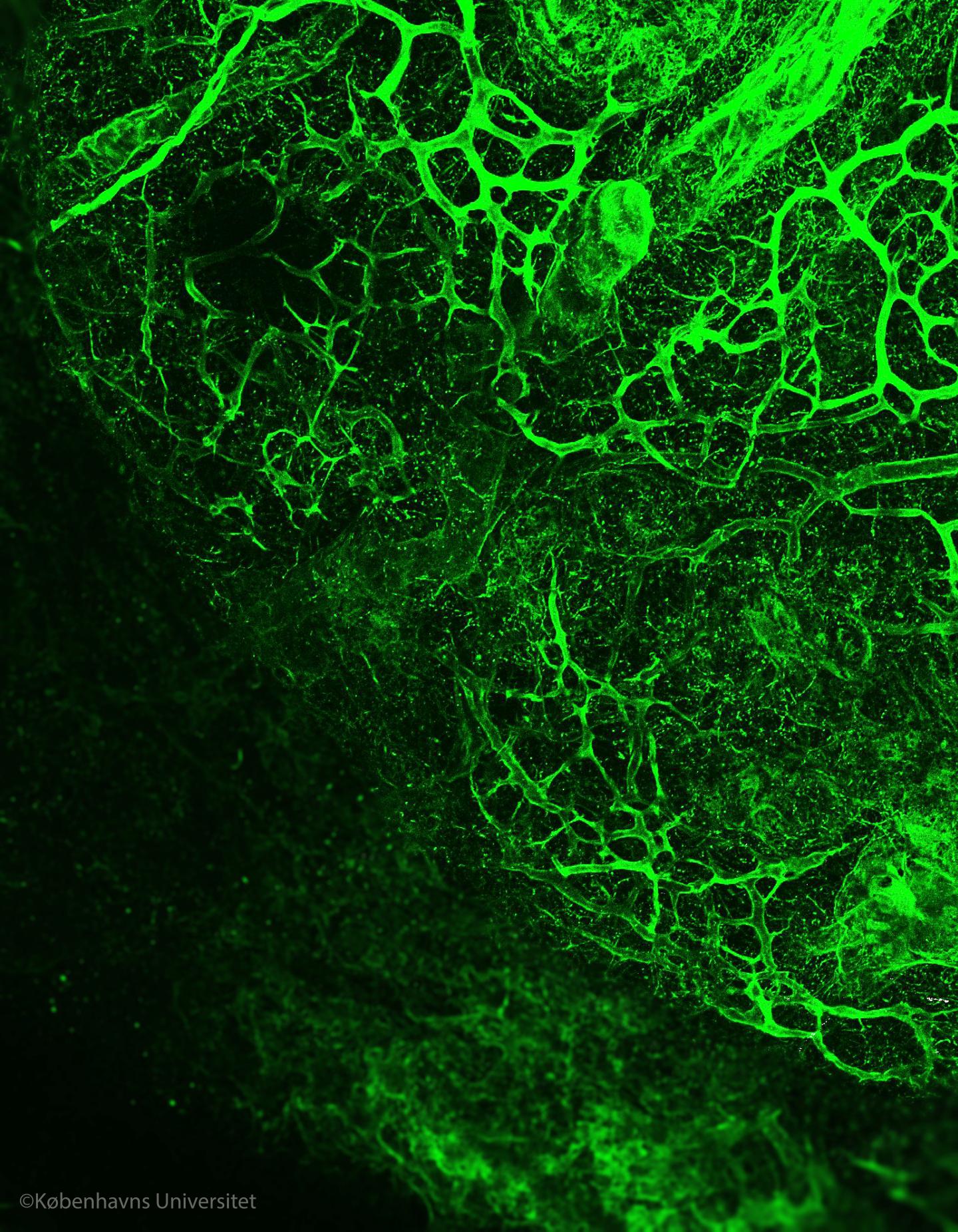

Credit: Image by Alejandro Mayorca-Guiliani.
- The matrix surrounds the cells in every organ of our bodies, and provides shape and structure to the organ. The matrix has a profound impact on how cells behave, so drives the progression of diseases such as cancer.
- A team of researchers from the Biotech Research & Innovation Centre (BRIC) at the University of Copenhagen, led by Professor Janine Erler, has developed a new technique published in Nature Medicine this month, that reveals the inner structure of organs and tumours by removing cells but leaving the matrix completely unaltered.
- The three-dimensional structure of this matrix has never been seen in such detail before.
"We have developed a technique to obtain intact organ scaffolds and to image them using microscopes. We are the first to image the structures of primary and metastatic tumours as well as healthy organs in this way", says Prof Erler.
A world of details revealed?Cells which are organised together to form tissues rely on the extracellular matrix as a foundation for attachment, to arrange themselves properly and to sense how to behave when their environment changes. Sometimes this organisation goes wrong and cells grow into tumours. To destroy a tumour, it is essential to know both its structure and the foundation upon which it is built.
This new method was pioneered by postdoctoral fellow Dr Alejandro Mayorca-Guiliani, in Prof Erler's team, who says, "We have isolated the structure that keeps tissues in place and organises the cells inside them. We did this by using existing blood vessels to deliver cell-removing compounds directly to a specific tissue to remove all cells within an organ, while leaving behind an intact scaffold that we could analyse biochemically and microscopically, providing us with the first view of the structure of tumours."
Imaging expert and co-author Chris Madsen (now at Lund University, Sweden) says "When you remove the cells, the clarity of what you can see through the microscope is much better – you can see the fibres of the matrix more clearly and you can look much deeper into the tissue."
Matrix biology expert and co-author Thomas Cox (now based at the Garvan Institute, Sydney) says "Because we're removing the cells completely, we can use mass spectrometry to identify and study the components of the matrix – in normal tissue and in tumours – in great detail".
Understanding cancer progression
This research is an advance in the fields of both cancer research and bioengineering: by using the decellularised organs we can learn much more about how tumours and normal organs are built, and what their differences are. This new technique might even have an impact on organ regeneration and tissue engineering in the future.
"We are now re-introducing cells into our extracellular matrix scaffolds, bringing them back to life, to study how tumours form and how cancer progresses. This is extremely exciting and offers a unique opportunity to study how cells behave in their native environment," explains Prof Erler.
###
The research is supported by the Danish Cancer Society, an ERC Consolidator Award, the Novo Nordisk Foundation, a European Research Council Consolidator Award, the Ragnar Söderberg Foundation Sweden, Cancerfonden Sweden, the Innovation Foundation Denmark, the National Health and Medical Research Council (NHMRC) Australia and the Danish Council for Independent Research.
Find the published article here: http://www.nature.com/nm/journal/vaop/ncurrent/full/nm.4352.html
Media Contact
Helen Frost
[email protected]
45-42-67-78-83
http://healthsciences.ku.dk/
Original Source
http://www.bric.ku.dk/research-stories/researchers-develop-new-technique-to-unveil-matrix-inside-tissues-and-tumours/ http://dx.doi.org/10.1038/nm.4352
############
Story Source: Materials provided by Scienmag

