Timely and precise diagnosis of malaria, a life-threatening infection caused by Plasmodium parasites transmitted to humans through the bites of infected Anopheles mosquitoes, remains essential for curbing transmission and lowering global mortality rates. With an estimated two hundred million new clinical cases and hundreds of thousands of fatalities reported globally each year, reliable laboratory confirmation remains an indispensable cornerstone of disease control. To accelerate the development of automated diagnostic tools that reduce diagnostic burdens on healthcare systems, researchers have introduced the DMLMD I dataset in a newly published data descriptor. Unlike many earlier benchmark collections that primarily targeted binary cell-level classification to distinguish pre-cropped healthy red blood cells from infected ones, this new collection specifically provides bounding-box object detection annotations across five developmental and multiplicity stages of intraerythrocytic Plasmodium falciparum. The primary dataset comprises 631 high-resolution thin blood smear microscopic images, which were expanded through a systematic, reproducible multi-layer augmentation pipeline to produce a 9,314-image corpus designed for deep learning model training.
The developmental lifecycle of the malaria parasite within the human host establishes the biological complexity that automated computer vision systems must successfully navigate. Human malaria is caused by five distinct species within the genus Plasmodium, with Plasmodium falciparum and Plasmodium vivax representing the most prevalent causes of infection worldwide. When an infected mosquito injects sporozoites into the bloodstream, the parasites travel to hepatocytes during an initial pre-erythrocytic phase before developing into merozoites that invade circulating erythrocytes. Inside host red blood cells, Plasmodium falciparum undergoes a forty-eight-hour intraerythrocytic progression passing through sequential developmental stages, including early ring forms, growing trophozoites, and mature schizonts. The subsequent rupture of infected erythrocytes releases hemozoin into the bloodstream, triggering the classic paroxysms of fever and chills that characterize the disease. In heavy infections, individual erythrocytes frequently harbor multiple invasive parasites, producing multi-ring or multi-trophozoite configurations that require specialized localization algorithms capable of distinguishing separate parasite instances.
Conventional diagnostic methodologies face practical trade-offs that highlight the persistent demand for automated microscopic analysis. Examination of Giemsa-stained blood smears under light microscopy has long been regarded as the clinical gold standard due to its high diagnostic sensitivity, low direct operational costs, and capacity to identify Plasmodium species, calculate parasite densities, and monitor response to medical therapy. In standard practice, stained thick blood smears offer roughly eleven times greater sensitivity than thin smears, providing an advantageous method for rapid screening in patients with low parasite counts, whereas thin blood smears preserve detailed parasite morphology to enable granular species identification and stage evaluation. However, manual light microscopy is heavily constrained by the requirement for qualified microscopists, variability in smear preparation, and the risk of diagnostic oversight or fatigue. Rapid diagnostic tests offer an alternative for point-of-care settings, but they suffer from lower sensitivity around one hundred parasites per microliter, persistent antigen detection, and susceptibility to variable storage conditions.
Alternative diagnostic tools beyond conventional bright-field microscopy also present substantial logistical challenges that limit widespread implementation in resource-constrained regions. Polymerase chain reaction assays offer remarkable analytical sensitivity and precise species discrimination even when circulating parasitemia is exceptionally low, yet their reliance on expensive thermal cyclers, delicate molecular reagents, and specialized personnel makes them impractical for routine clinical diagnosis. Similarly, monochromatic flow cytometry systems that count infected cells and examine intraerythrocytic development through laser scattering operate effectively in high-end experimental facilities but cannot easily be adapted to field settings. Serological evaluations such as enzyme-linked immunosorbent assays and immunofluorescence antibody tests detect circulating antibodies or specific diagnostic proteins like histidine-rich protein 2, but they struggle to differentiate acute infections from past exposure. These persistent bottlenecks underscore the necessity of developing scalable, computer-assisted optical screening platforms that can automatically inspect standard Giemsa-stained microscopic fields without requiring costly physical infrastructure.
In response to these clinical demands, researchers have increasingly deployed computer vision and deep learning architectures to analyze digitized microscopy slides. Early computational experiments evaluated blood smear images across varying optical magnifications, and subsequent investigations utilized convolutional neural networks, Mask R-CNN architectures, and custom object detectors such as YOLO variants and transformer-based AIDMAN models to detect or segment infected erythrocytes. Despite achieving reported diagnostic accuracies exceeding ninety-four to ninety-eight percent in controlled evaluations, many existing models remain constrained by the narrow scope of public training datasets. Many historical repositories only provide individual, pre-cropped single cells categorized along binary lines as infected or uninfected, completely omitting broader contextual field features. Furthermore, many existing collections lack adequate morphological coverage of diverse parasite stages or fail to document complex multi-infection occurrences, which limits their generalization when applied to real-world microscopic fields containing crowded cell populations and variable parasitemia levels.
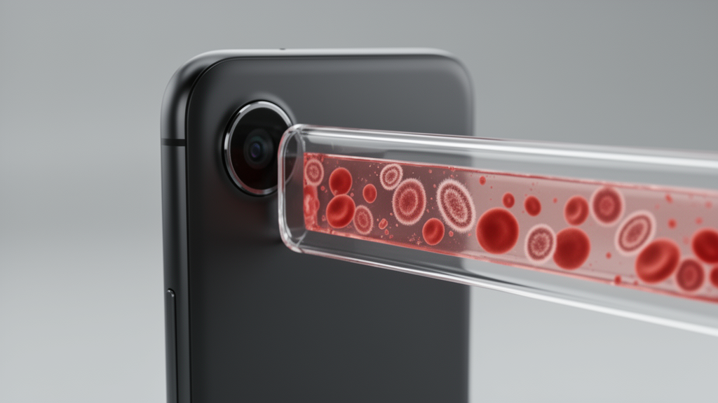
The DMLMD I repository directly addresses these diagnostic limitations by providing multi-object, multi-class bounding-box annotations derived from authentic thin blood smear preparations. Over multiple culture batches of the Plasmodium falciparum 3D7 strain prepared between 2023 and 2024, laboratory personnel created 250 thin blood smears under controlled Biosafety Level II conditions. High-magnification microscopic fields were systematically acquired at one-thousand-times optical magnification using a forty-eight-megapixel smartphone camera coupled via an optical adaptor to a conventional bright-field microscope equipped with a one-hundred-times oil-immersion objective and Kohler illumination. To maximize optical consistency, exposure parameters and white balance settings were maintained throughout imaging sessions. A dedicated malaria specialist established strict quality control filtering, screening captured fields to exclude visual artifacts such as optical blurring, dense cellular stacking, and dye precipitation. From this initial material, 631 high-resolution fields showing evenly distributed red blood cell monolayers were selected as the primary unaugmented benchmark dataset.
Annotation procedures for DMLMD I were structured specifically for object localization rather than basic whole-image categorization. For every observable parasite instance, a tight bounding box was drawn around the boundary of the host erythrocyte, capturing five distinct infection categories: Ring stage, Trophozoite stage, Schizont stage, Multi Ring stage, and Multi Trophozoite stage. Because individual fields of view frequently present multiple parasites or coexisting developmental phases, the cumulative instance count significantly exceeds the total image count, establishing a rich multi-class distribution. To eliminate individual annotator bias and verify spatial precision, a systematic audit evaluated a randomly chosen fifteen-percent validation subset comprising ninety-five images and 358 infected erythrocyte instances. A second senior parasitologist independently re-annotated this verification cohort. Using an intersection over union threshold of at least 0.50 to confirm spatial bounding-box overlap, the independent cross-evaluation confirmed a 95.53 percent spatial localization agreement across 342 matched instances, with all remaining discrepancies resolved through consensus adjudication before dataset finalization.
To provide sufficient geometric and optical variability for training modern deep learning frameworks, the researchers established a reproducible four-layer data augmentation pipeline using Roboflow. The baseline repository of 631 expert-annotated primary images was sequentially processed through four cumulative operational layers. The initial layer applied horizontal mirroring to double geometric orientations, followed by a second layer incorporating vertical flips to emulate camera and slide positional shifts. A third layer introduced ninety-degree rotational steps to instill rotational invariance across diverse slide placements, while the fourth layer injected a one-percent noise perturbation to simulate microscopic background grain, camera sensor noise, and compression artifacts. Through this cumulative protocol, the collection expanded from the baseline 631 images to a final verified volume of 9,314 annotated images. Every generated image retains an individual metadata token linking back to its original parent image index, preserving complete biological provenance and preventing inadvertent data duplication across training workflows.
Analysis of the annotated labels reveals crucial insights into class balance and spatial distribution across the augmented corpus. Within the dataset, single trophozoite forms represent the most frequent class, while multi-ring and multi-trophozoite annotations capture dense infection events that reflect elevated parasitemia in clinical blood samples. Spatial coordinate evaluations demonstrate that annotated bounding boxes exhibit a central clustering tendency across field dimensions, reflecting standard microscopist framing practices, while bounding-box width and height distributions indicate consistent red blood cell diameters alongside larger boundaries corresponding to mature schizonts or multi-parasite clusters. The investigators explicitly emphasized proper data hygiene for downstream computational research: users must divide data at the source image level prior to augmentation, ensuring that holdout test and validation partitions contain solely unaugmented base images from isolated slides. This rigorous separation prevents feature leakage between evaluation sets and guarantees that measured neural network performance accurately reflects real generalization capability.
Looking ahead, the authors position DMLMD I as a foundational resource for building scalable, point-of-care artificial intelligence systems while recognizing important boundaries of the current release. Because the baseline images originate from in vitro cultures of the standardized Plasmodium falciparum 3D7 strain, they do not encompass the full morphological heterogeneity seen in field clinical smears, where patient erythrocyte anomalies, co-infecting non-falciparum species, variable staining chemistries, and erratic illumination conditions are common. The systematic inclusion of geometric rotations, flips, and sensor noise aims to bridge part of this domain shift, but prospective clinical validation and testing across diverse neural architectures will be necessary before bedside deployment. By placing multi-class intraerythrocytic stage detection and multiplicity at the core of an open-access image repository, the study provides machine learning engineers and parasitologists with an annotated benchmark to train real-time, smartphone-compatible diagnostic models capable of assisting overworked healthcare workers across resource-constrained endemic areas.
Subject of Research: Computer vision dataset for automated object detection of malaria parasite stages
Article Title: DMLMD I dataset for machine learning based malaria detection using blood smears
Article References: Sharma, A., Jangra, R., Deepa, Yadav, Y., Badhwar, P., Joshi, K., Singh, S., Gill, S. S., & Gill, R. (2026). DMLMD I dataset for machine learning based malaria detection using blood smears. Discover Informatics, 1(1), Article 36. https://doi.org/10.1007/s44564-026-00036-9
Image Credits: AI Generated
DOI: 10.1007/s44564-026-00036-9
Keywords: Malaria Detection, Deep Learning, Computer Vision, Blood Smears, Plasmodium falciparum, Object Detection, Roboflow, Microscopy, DMLMD, dataset, machine, learning
News Source: Blake Davidson. (November 9, 2026). New Open Dataset Uses Smartphone Microscopy to Power AI Malaria Detection. Scienmag.