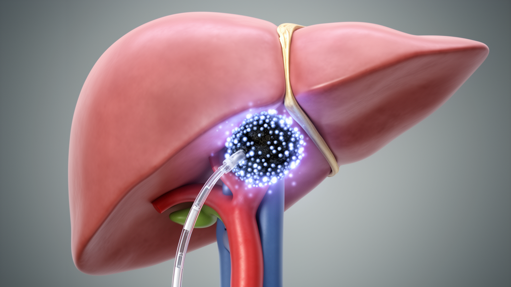
Researchers have demonstrated the feasibility of a novel computational framework designed to optimize treatment plans for transarterial radioembolization (TARE) in patients with hepatocellular carcinoma. The study, published in EJNMMI Physics, introduces a patient-specific modeling approach that allows for the systematic optimization of specific activity to maximize tumor absorbed dose while minimizing radiation exposure to healthy liver tissue. This work represents a significant step toward personalized dosimetry strategies, addressing the growing need for predictive methods as TARE evolves from a palliative to a curative treatment modality for liver cancer.
The research team, led by Carlos Huesa-Berral and Alejandro Bertolet, developed a framework named MIDOS to design optimal TARE plans. The study was conducted as a retrospective cohort analysis involving fourteen patients with solitary hepatocellular carcinoma who had previously undergone [99mTc]Tc-MAA SPECT-CT imaging. The cohort included both segmental and lobar administrations, providing a range of clinical scenarios for the evaluation of the modeling framework. By analyzing this specific group of patients, the researchers aimed to assess the practical applicability of their proposed optimization method in real-world clinical contexts.
The core of the MIDOS framework involves the integration of patient-specific anatomical and physiological data into a liver vasculature model. For each patient in the cohort, segmentations of the tumor and the perfused area were incorporated into the model. The simulation process assigned microspheres stochastically in cluster patterns to either tumor or normal liver tissue. The probabilities of these assignments were proportional to blood flow and were further modulated by specific uptake models for both tumor and normal liver tissues. This detailed simulation allowed for the generation of synthetic [99mTc]Tc-MAA SPECT images that could be directly compared with the actual patient images to validate the accuracy of the model.
To validate the synthetic images, the researchers employed several quantitative metrics, including relative activity errors in tumor and normal tissue, the tumor-to-normal ratio (TNR), and gamma analysis of normal tissue distribution. The results indicated high fidelity in the modeling process. Synthetic [99mTc]Tc-MAA SPECT images presented relative errors below 2% for overall tumor activity, normal liver activity, and the TNR. Furthermore, the pass rates for the 10%/10 mm gamma analysis of normal tissue distribution exceeded 90% in all patients. These findings suggest that the MIDOS framework can accurately replicate the physical distribution of radiopharmaceuticals within the liver, providing a reliable basis for subsequent treatment planning.
Using the validated simulations, the team generated virtual Y-90 treatment plans across a wide range of specific activities, from 40 to 4000 Bq/bead. From these plans, personalized treatment planning curves were derived, illustrating the relationship between specific activity and key dosimetric parameters. These curves included the TNR, as well as the mean absorbed dose to both the tumor and the normal liver. The analysis revealed that the optimal specific activity varied significantly between patients and clinical scenarios.
The treatment planning curves identified patient- and scenario-specific optima in the TNR, which corresponded to the maximal tumor mean dose and the minimal normal liver mean dose. The study demonstrates that the MIDOS framework can systematically identify these optimal points, providing a quantitative tool for treatment decision-making.
The framework’s capacity to handle different clinical scenarios, such as segmental versus lobar administrations, makes it a versatile tool for various patient presentations. The study confirms that quantitative treatment optimization is feasible and can be integrated into the planning workflow for TARE.
Despite the promising results, the study has certain limitations inherent to its retrospective design and the specific cohort size. The analysis was limited to fourteen patients with solitary HCC, which may not fully represent the diversity of liver cancer cases encountered in clinical practice. The use of synthetic images for validation, while showing high accuracy, relies on the assumptions of the underlying uptake models. Nevertheless, this research provides a robust foundation for the development of patient-specific optimization tools in transarterial radioembolization, potentially improving treatment outcomes for patients with hepatocellular carcinoma.
Subject of Research: Optimization of transarterial radioembolization treatment plans using patient-specific modeling for hepatocellular carcinoma.
Article Title: Patient-specific modeling enables specific activity optimization to maximize tumor absorbed dose in transarterial radioembolization
Article References: Huesa-Berral, C., Dawson, R. J., Sabet, H., Rodríguez-Fraile, M., Atta, R., Paganetti, H., Bolch, W. E., Wehrenberg-Klee, E., & Bertolet, A. (2026). Patient-specific modeling enables specific activity optimization to maximize tumor absorbed dose in transarterial radioembolization. EJNMMI Physics. https://doi.org/10.1186/s40658-026-00921-0
Image Credits: AI Generated
DOI: 10.1186/s40658-026-00921-0
Keywords: transarterial radioembolization, hepatocellular carcinoma, patient-specific modeling, dosimetry, Y-90 microspheres, treatment planning, nuclear medicine, liver cancer, Patient-specific, modeling, enables, specific
News Source: Nathaniel Bowman. (November 10, 2026). New Model Optimizes Radioembolization Dose for Liver Cancer. Scienmag.