Mesonephric carcinoma and mesonephric-like adenocarcinoma represent rare but aggressive malignancies within the female genital tract, captivating the attention of pathologists and oncologists due to their unusual origins, complex morphology, and challenging diagnosis. These neoplasms defy conventional categorization, exhibiting unique histopathological and molecular features that set them apart from the more commonly encountered gynecologic cancers. A deeper understanding of their biology not only advances diagnostic precision but also paves the way for more tailored therapeutic strategies.
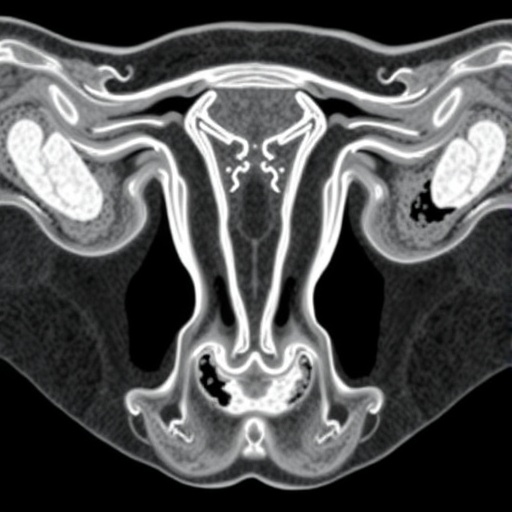
At the core of mesonephric carcinoma lies its origin from mesonephric remnants—vestigial structures derived from the Wolffian duct system predominantly located in the lateral wall of the cervix. These remnants can persist into adulthood as benign cellular aggregates or undergo hyperplastic changes. Mesonephric carcinoma arises when these remnants undergo malignant transformation, leading to adenocarcinomas marked by a constellation of diverse architectural patterns, including tubular, papillary, and solid growths. The presence of nuclear features reminiscent of papillary thyroid carcinoma adds another layer of diagnostic complexity.
Histologically, normal mesonephric remnants are characterized by small, tightly packed tubules lined by cuboidal epithelium containing eosinophilic secretions but lacking cilia and mucin production. This subtle architectural pattern can be mistaken for benign processes, especially when proliferative changes such as mesonephric hyperplasia occur. Differentiating benign hyperplasia from frank carcinoma demands keen histopathological scrutiny combined with immunohistochemical profiling, underscoring the clinical importance of recognizing the unique biomarker expression of these lesions.
Immunohistochemistry serves as a fundamental tool in the diagnostic armamentarium. Mesonephric carcinoma typically exhibits strong positivity for transcription factors including PAX8 and GATA3, with luminal expression of CD10 and focal positivity for TTF1. This immunoprofile is distinct with consistent negativity for estrogen receptor (ER), progesterone receptor (PR), and p16—markers commonly expressed in other gynecologic adenocarcinomas. Additionally, p53 usually displays a wild-type staining pattern, and the mismatch repair (MMR) proteins remain intact, delineating mesonephric carcinoma from more common tumor subtypes driven by these pathways.
From a molecular perspective, mesonephric carcinoma reveals nearly universal mutations in the KRAS or NRAS oncogenes, genetic alterations that activate key signaling pathways driving tumorigenesis. Chromosomal aberrations frequently observed include gains of chromosome arm 1q and losses of 1p, coupled with mutations in chromatin remodeling genes such as ARID1A, ARID1B, and SMARCA4. Notably absent are mutations commonly seen in Müllerian-derived endometrioid carcinomas, especially those affecting PIK3CA and PTEN, highlighting a distinct oncogenic trajectory.
Contrasting with mesonephric carcinoma, mesonephric-like adenocarcinoma (MLA) emerges primarily in the endometrium and ovary, without clear association with recognizable mesonephric remnants. Despite morphologic similarity to mesonephric carcinoma, MLA is thought to derive from Müllerian epithelium that exhibits divergent mesonephric differentiation. This hypothesis is reinforced by frequent coexistence with Müllerian precursor lesions such as endometriosis and atypical hyperplasia, placing MLA within a unique pathogenetic niche.
Immunohistochemical evaluation of MLA reveals overlapping yet distinguishable features from mesonephric carcinoma. While it commonly demonstrates diffuse positivity for TTF1, GATA3 expression is often weaker and more variable. The molecular profile overlaps with mesonephric carcinoma, sharing KRAS or NRAS mutations as well as chromosomal gains and losses; however, MLA also harbors mutations in PIK3CA, PTEN, or CTNNB1. These latter alterations align MLA more closely with Müllerian neoplasms and underscore its divergent molecular evolution compared to true mesonephric carcinoma.
The diagnostic challenge posed by MC and MLA is compounded by their histological heterogeneity, which necessitates careful differential diagnosis against other aggressive gynecologic tumors. Endometrioid adenocarcinoma, frequently ER/PR-positive and occasionally exhibiting squamous or mucinous differentiation, must be distinguished through immunoprofiling. Clear cell carcinoma presents a separate morphological and immunophenotypic entity marked by Napsin A, HNF1-beta, and AMACR positivity, whereas high-grade serous carcinoma harbors TP53 mutations and dramatic nuclear atypia.
Rare adnexal tumors such as Female Adnexal Tumor of Probable Wolffian Origin (FATWO) and stromal tumors with STK11 mutations further confound the diagnostic landscape. FATWO lacks PAX8 expression, a marker robustly expressed in both MC and MLA, while STK11-mutated tumors possess distinct molecular signatures. These nuanced distinctions underscore the imperative of integrated histopathological and molecular assessments in the accurate classification of these tumors.
Clinically, both mesonephric carcinoma and mesonephric-like adenocarcinoma follow an aggressive course with a notable propensity for distant metastases. The rarity of these tumors and their overlapping yet non-identical features often lead to diagnostic delays or misclassification, potentially impacting patient outcomes adversely. As such, heightened clinical awareness and implementation of advanced diagnostic modalities are critical for early detection and intervention.
The emerging molecular landscape not only informs diagnosis but also unlocks new avenues for targeted therapy. The ubiquity of KRAS/NRAS mutations highlights the potential role of inhibitors targeting the RAS-RAF-MEK-ERK pathway, although clinical trials remain in early stages. Meanwhile, the presence of chromatin remodeling gene mutations invites exploration of epigenetic therapies tailored to these alterations. Understanding the distinctions between MC and MLA at the molecular level will be pivotal in developing personalized management strategies.
Overall, mesonephric carcinoma and mesonephric-like adenocarcinoma exemplify the evolving complexity of gynecologic oncology, where traditional histology alone cannot suffice. The integration of histopathology, immunophenotyping, and molecular characterization embodies the future of pathologic classification, enabling precise tumor identification and tailored therapeutic approaches. The expanding body of research continues to reveal the intricate interplay between embryological origin, molecular alterations, and tumor behavior in these enigmatic neoplasms.
Continued multidisciplinary collaboration between pathologists, molecular biologists, and clinicians will be essential to unravel the full spectrum of mesonephric and mesonephric-like tumors. Prospective studies elucidating prognostic factors and treatment responses are urgently needed to translate laboratory findings into improved patient care. These rare tumors highlight not only scientific challenges but also the promises of precision medicine in gynecologic cancer.
In conclusion, mesonephric carcinoma and mesonephric-like adenocarcinoma stand at the crossroads of developmental biology and oncogenesis, their distinct origins reflected in their pathology and genetics. Diagnostic vigilance combined with advanced molecular insights can empower clinicians in confronting these aggressive malignancies, offering hope for more effective interventions and better patient outcomes in the years ahead.
Subject of Research:
Mesonephric carcinoma and mesonephric-like adenocarcinoma of the female genital tract
Article Title:
Mesonephric Carcinoma and Mesonephric-like Adenocarcinoma of the Female Genital Tract
News Publication Date:
14-Jul-2025
Web References:
http://dx.doi.org/10.14218/JCTP.2025.00020
Keywords:
Immunohistochemistry, Molecular biology, KRAS mutations, Gynecologic oncology, Mesonephric carcinoma, Mesonephric-like adenocarcinoma, PAX8, GATA3, TTF1, Chromatin remodeling, Müllerian differentiation
Tags: architectural patterns in adenocarcinomasdiagnostic challenges in mesonephric carcinomahistopathological features of mesonephric tumorshyperplastic changes in mesonephric tissuemesonephric carcinoma in female genital tractmesonephric-like adenocarcinoma characteristicsmolecular biology of mesonephric neoplasmsorigins of mesonephric remnantspathologists and oncologists insightsrare gynecologic cancers diagnosistherapeutic strategies for rare cancersunique


