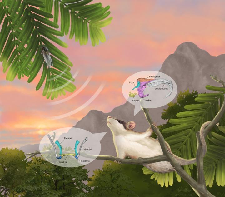
Credit: IVPP
A joint research team led by Dr. MAO Fangyuan from the Institute of Vertebrate Paleontology and Paleoanthropology (IVPP) of the Chinese Academy of Sciences and Prof. MENG Jin from the American Museum of Natural History has reported a new multituberculate mammal, Sinobaatar pani, with well-preserved middle ear bones.
The new mammal comes from the Early Cretaceous Jehol Biota in Northeast China. Comparing three types of fossils with extant mammals at different embryological stages, the researchers identified various evolutionary stages and ancestral phenotypes of the mammalian middle ear.
Their findings were published in National Science Review on Aug. 25.
For mammals, the external ear (the pinna) collects airborne sounds that vibrate the eardrums, and the middle ear bones on the inner side of the eardrum function as a delivery system that transmits sound vibrations to the inner ear.
According to previous studies, we know that the extra mammalian ear bones actually originated from the jawbones of reptiles. However, few studies have actually looked at the detailed morphologies of ear bones that are the ancestral phenotypes for the middle ear of modern mammals.
Multituberculates are an extinct group of mammals that lived from the Middle Jurassic (about 165 million years ago) to the Eocene (about 35 million years ago). The most exciting discovery about the new animal is its middle ear bones, which are the first unequivocal evidence of the five auditory bones from this extinct mammalian group.
These miniscule bones are still embedded in rock and not visible. Using computerized tomography (CT), MAO and her colleagues were able to digitally “extract” the ear bones from the rock and reconstruct them in three-dimensional form so that their morphology could be observed in detail.
The data provided by MAO and her colleagues are by far the best evidence of middle ear morphology in known Mesozoic mammals. For comparison, the data also included similar CT reconstructions of the middle ear of extant monotremes, marsupials and placentals.
“There are two basic patterns of the middle ear in living mammals, represented by monotremes and therians, respectively. In the former, the middle ear is characterized by an ‘abutting contact’ between the incus and malleus, which is distinct from the one in therian mammals where the incus-malleus articulation is saddle-shaped,” said Dr. MAO.
The researchers recognized that the three main Mesozoic mammalian groups (i.e., multituberculates, eutriconodontans, and symmetrodontans) share a similar middle ear structure between the incus and malleus, which they termed the “braced hinge joint”.
Although they acknowledged that the middle ear may have evolved independently in several mammalian groups, they proposed that the braced hinge joint could represent a critical feature of the ancestral phenotype of the mammalian middle ear.
The abutting pattern in monotremes and the saddle-shaped joint in therians may well be derived from the braced hinge joint linking the incus and malleus as observed in Mesozoic mammals. At the least, these fossil forms have narrowed the morphological gap between the middle ear of mammal-like reptiles, formed by the postdentary bones lodged in the lower jaw, to the middle ear of extant mammals.
The researchers proposed that the surangular bone, which is another postdentary bone in mammal-like reptiles, persisted in Mesozoic mammals; its fate in living mammals remains uncertain.
They further showed that middle ear morphologies in Mesozoic mammals represent different evolutionary stages, with that of Liaocodondon being the most primitive, Origolestes the intermediate, and Sinobaatar the most advanced.
Developmental features observed in extant mammals and evolution of the mammalian middle ear in fossil records are correlated, said MAO.
###
Media Contact
MAO Fangyuan
[email protected]
Related Journal Article
http://dx.


