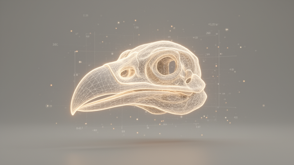
Researchers have developed a fully unsupervised geometric framework designed to extract robust parametric descriptors of cranial morphology directly from imperfect 3D skull meshes, bypassing the need for labor-intensive manual landmarking. This advancement addresses a significant bottleneck in comparative biology, where existing high-throughput analysis pipelines have often been hindered by the requirement for high-quality data and human intervention. By leveraging computational geometry, differential geometry, and advanced numerical optimization, the new method automatically aligns specimens and measures primary skull dimensions, ensuring stability even when processing topologically noisy or damaged digital collections.
The study validated the automated pipeline using 100 skull specimens representing Darwin’s finches and their relatives. The framework successfully recovered the well-established scaling relationship between skull size and orbit curvature, confirming the biological accuracy of its measurements. Building on these findings, the team established a predictive model capable of estimating orbit curvature from three easily obtainable linear skull measurements. This model explained 86.36% of the variance in curvature with an average prediction error of only 6.14%, demonstrating the precision of the derived parameters.
In addition to these predictive capabilities, the researchers derived dimensionless relative-geometry descriptors that remain invariant to uniform scaling while retaining meaningful variation among the sampled genera. The generalizability of this method was further confirmed through successful applications to a broader range of avian and mammalian skulls. These results suggest that the framework provides a rigorous, scalable foundation for digital estimation and high-throughput phenotyping of large-scale museum collections.
By automating the extraction of morphometric data from imperfect 3D scans, this geometric framework facilitates the study of large, complex anatomical datasets that were previously difficult to analyze efficiently. The ability to generate stable, parametric descriptors without manual input represents a major step forward for researchers aiming to quantify cranial morphology at scale. This development could substantially accelerate comparative biological research, allowing for more consistent and rapid analysis of diverse evolutionary specimens.
Subject of Research: Computational morphology and automated 3D analysis in comparative biology
Article Title: Robust parametric estimation of avian cranial morphology
Article References: Lau, K., & Choi, G. P. T. (2026). Robust parametric estimation of avian cranial morphology. PLOS Computational Biology, 22(11), e1014941. https://doi.org/10.1371/journal.pcbi.1014941
Image Credits: AI Generated
DOI: 10.1371/journal.pcbi.1014941
Keywords: cranial morphology, 3D imaging, computational geometry, comparative biology, Darwin's finches, automated phenotyping, morphometrics, geometric primitives, numerical optimization, skull measurement, orbit curvature, predictive modeling, museum collections, digital evolution, bioinformatics, unsupervised learning, avian anatomy, mammalian skulls
News Source: Drew Townsend. (November 16, 2026). New Automated Framework Accurately Estimates Avian Cranial Morphology from 3D Scans. Scienmag.