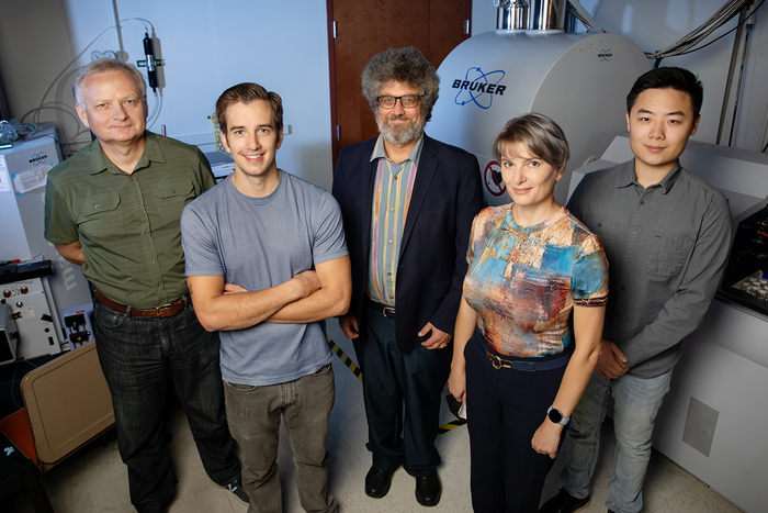
CHAMPAIGN, Ill. — Researchers at the University of Illinois Urbana-Champaign can now rapidly isolate and chemically characterize individual organelles within cells. The new technique tests the limits of analytical chemistry and rapidly reveals the chemical composition of organelles that control biological growth, development and disease.

Credit: Photo by L. Brian Stauffer
CHAMPAIGN, Ill. — Researchers at the University of Illinois Urbana-Champaign can now rapidly isolate and chemically characterize individual organelles within cells. The new technique tests the limits of analytical chemistry and rapidly reveals the chemical composition of organelles that control biological growth, development and disease.
The findings of the new study, led by chemistry professor Jonathan Sweedler, are published in the journal Nature Methods.
The new approach locates and isolates individual organelles using light microscopy, then chemically analyzes them via MALDI MS, or matrix-assisted laser desorption/ionization mass spectrometry. The entire process takes an hour – a task that could take human analysts years to complete.
“Cells are not just little sacks full of chemicals,” Sweedler said. “They contain organelles that perform specific functions. The ability to characterize the chemical composition of individual organelles should lead to a better understanding of how cells develop and express diseases.”
The researchers said they are not the first to characterize organelles chemically. But using their automated targeting and chemical analysis approach is faster and more accurate, and assures that they analyze exactly what they intend. This way, they can determine the chemical makeup of a single organelle – not the average composition of a larger sample containing many organelles.
For this study, the team focused on the cell’s vesicles – both dense-core and lucent varieties – collected from sea slugs, which are a commonly used neuroscience study model. Vesicles were selected as the organelle of interest because they are involved in chemical cell-to-cell signaling. The researchers said they are also larger than many of the other organelles, making them excellent first candidates to demonstrate the capabilities of the new approach.
“We analyzed approximately 1,000 individual vesicles from sea slugs,” chemistry professor Stanislav Rubakhin said. “We found heterogeneity among the types of lipids and biologically active peptides, indicating that MALDI MS is sensitive enough to detect chemical differences between what were thought to be the same types of organelles.”
Because disease is often spotted when heterogenous cells appear within a single tissue type, Rubakhin said, the ability to discern these differences at the subcellular level could lead to earlier detection and treatment.
“Our new workflow can help the scientific community complete the ‘parts list’ of the organelles found within cells,” graduate student Daniel Castro said. “Having that parts list will help us determine if something is missing or extra within the organelles, helping us spot subtle changes and study how those changes correlate to diseases such cancer and those related to the brain and mental health.”
The National Institute on Drug Abuse and the National Human Genome Research Institute supported this study.
Sweedler is the director of the School of Chemical Sciences and is affiliated with the Beckman Institute for Advanced Science and Technology, the Carl R. Woese Institute for Genomic Biology and the Carle Illinois College of Medicine.
Editor’s notes:
To reach Jonathan Sweedler, call 217-244-7359; email [email protected].
The paper “Image-guided MALDI mass spectrometry for high-throughput single- organelle characterization” is available online and from the U. of I. News Bureau.
DOI: 10.1038/s41592-021-01277-2.
Journal
Nature Methods
DOI
10.1038/s41592-021-01277-2
Method of Research
Observational study
Subject of Research
Cells
Article Title
Image-guided MALDI mass spectrometry for high-throughput single- organelle characterization
Article Publication Date
30-Sep-2021
COI Statement
The authors declare no competing interests.


