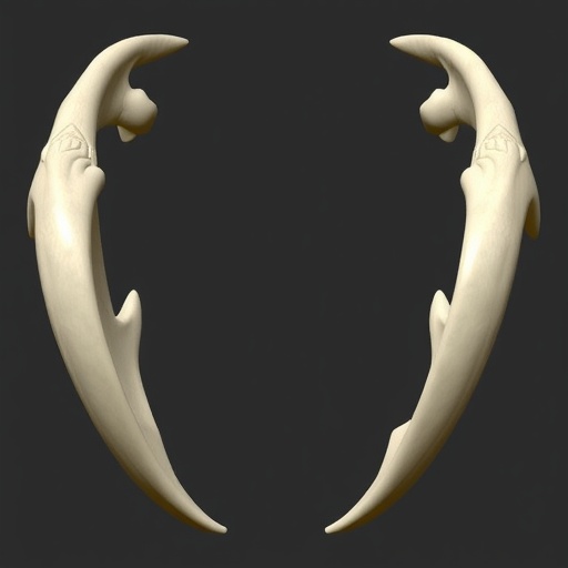
In the ever-evolving realm of forensic science and medical anthropology, reconstructing missing skeletal elements has long posed a formidable challenge. Among these, the mandible—the lower jawbone—holds a pivotal role, not only in facial structure and identity but also in forensic identification and trauma analysis. Recognizing this critical need, a pioneering study has emerged that introduces an innovative and highly precise approach to estimate missing mandibular structures leveraging three-dimensional homologous modeling techniques. This breakthrough, detailed in the recent publication by Namiki, Makino, Iwase, and colleagues, marks a significant leap forward in forensic methodologies, potentially revolutionizing the way forensic experts and anthropologists tackle incomplete skeletal remains.
Traditional approaches to mandible reconstruction often involve direct morphometric comparisons, manual sculpting, or extrapolations based on fragmented remains and comparative anatomical data. These methods, while valuable, are frequently limited by subjectivity, insufficient anatomical precision, and, at times, the inability to account for individual variability. Namiki et al.’s novel method seeks to overcome these constraints by utilizing highly sophisticated three-dimensional homologous models, which provide a robust, mathematically grounded framework for reconstructing missing bone structures with unprecedented accuracy.
The innovation hinges on the concept of homologous landmarks—specific anatomical reference points that are consistent across different human mandibles despite individual morphological differences. By compiling extensive 3D datasets of these landmarks from diverse samples, the researchers constructed a comprehensive homologous model that embodies the spectrum of mandibular shape variations. This model then serves as the baseline for estimating the geometry of absent mandibular segments in forensic samples, effectively “filling in the gaps” based on statistically validated morphometric correlations.
One of the remarkable aspects of this methodology is its reliance on three-dimensional computational techniques, which allow for an exhaustive comparison across multiple dimensions rather than simplistic two-dimensional projections. This grants the model enhanced adaptability when confronting partial or severely damaged mandibles, which are commonplace in forensic contexts. Additionally, this digital framework ensures repeatability and objectivity, reducing the risks associated with manual estimations.
The potential applications of this method go beyond forensic casework. In clinical settings, the accurate reconstruction of mandibular defects is crucial for surgical planning, prosthetic design, and rehabilitation after trauma or tumor resections. The integration of 3D homologous models could substantially improve the precision of maxillofacial surgeries by enabling surgeons to anticipate anatomical contours and functionalities that are lost or altered. Furthermore, research in human evolution and population biology could benefit from such advanced morphometric tools to better understand variations and developmental processes in mandible morphology.
Furthermore, the computational nature of the novel technique invites future expansions, such as integration with machine learning algorithms. By training AI networks on extensive mandibular datasets, these systems could predict missing structures with increasing accuracy and efficiency, potentially automating aspects of forensic reconstruction that currently require expert intervention. As digitized anatomical archives grow, these models could evolve into adaptive platforms, continually refining their reconstruction capabilities.
Importantly, Namiki and collaborators validated their approach through rigorous testing on mandibles with artificially introduced defects, demonstrating that their method reliably recovers missing anatomical features. This validation is critical, as forensic reconstructions must withstand legal scrutiny and provide dependable data for identification or investigative purposes. The ability to present a scientifically robust reconstruction can directly impact judicial outcomes, victim identification, and the resolution of cases involving skeletal remains.
Moreover, the research represents a synthesis of multiple disciplines, spanning forensic medicine, computer science, anatomy, and statistical shape analysis. This interdisciplinary collaboration exemplifies how modern science transcends traditional boundaries, employing computational power and biological insight to solve complex practical problems. The study also highlights the importance of open, comprehensive anatomical databases that serve as repositories for continued refinement of homologous modeling.
While promising, the implementation of this technique requires access to advanced imaging technologies such as CT and laser scanning to generate accurate 3D models of forensic samples. This may present logistical and financial challenges, particularly in regions with limited forensic infrastructure. Nonetheless, the increasing availability and decreasing costs of 3D scanning technologies suggest that such barriers will diminish over time, making the method increasingly accessible worldwide.
Ethical considerations also arise, especially concerning the handling of human remains and privacy in the creation and use of anatomical databases. The research community must address these concerns by establishing guidelines that respect the dignity and cultural sensitivities associated with human skeletal materials. Transparent protocols and informed consent procedures will be essential to foster trust and cooperation among stakeholders.
In addition to ethical and infrastructural aspects, the method’s applicability across different populations remains an area for further investigation. Mandibular morphology exhibits significant variation influenced by genetic, environmental, and cultural factors. Therefore, expanding the homologous model datasets to include diverse populations will ensure that the technique remains valid and universally applicable, avoiding biases and improving forensic accuracy globally.
The implications of this advancement also resonate in educational contexts. Forensic anthropology students and professionals could employ such 3D models as teaching aids, giving learners hands-on experience with a wide variety of anatomical forms and pathologies without relying solely on physical specimens. This digital democratization of anatomical reference material enhances training quality and fosters innovation in forensic sciences.
Finally, this developmental work signals a growing trend towards digitization and computational analysis in medical and forensic sciences—a paradigm shift that leverages artificial intelligence, 3D modeling, and big data to enhance precision and reliability. As forensic investigations become increasingly complex, methods like the one proposed by Namiki et al. become indispensable tools, augmenting human expertise through technology.
In conclusion, the newly developed method for estimating missing mandibles using three-dimensional homologous models represents an exciting frontier in forensic medicine and beyond. By combining anatomical rigor, computational sophistication, and interdisciplinary collaboration, this technique offers a scalable, objective, and highly accurate solution to a longstanding challenge. As the approach gains traction and evolves, it promises to transform forensic reconstruction practices, offering more reliable victim identification and contributing to justice served with scientific integrity.
Subject of Research: Forensic reconstruction of missing mandibular bone structures using three-dimensional homologous models.
Article Title: Development of a novel method for estimating the missing mandible using three-dimensional homologous models.
Article References:
Namiki, S., Makino, Y., Iwase, H. et al. Development of a novel method for estimating the missing mandible using three-dimensional homologous models. Int J Legal Med (2026). https://doi.org/10.1007/s00414-025-03711-y
DOI: https://doi.org/10.1007/s00414-025-03711-y
Tags: 3D modeling in anthropologyadvanced morphometric techniquesanatomical precision in forensicsestimating missing mandiblesforensic science techniqueshomologous landmarks in anthropologyindividual variability in skeletal remainsinnovative forensic methodologiesmandibular structure analysisrevolutionizing forensic anthropologyskeletal reconstruction methodstrauma analysis and identification


