In a groundbreaking development poised to redefine the early diagnostic landscape of Parkinson’s disease (PD), a recent comparative MRI study has demonstrated that neuromelanin imaging significantly outperforms free water imaging in detecting the disease at its nascent stages. Published in the highly regarded journal npj Parkinsons Dis. in 2026, this study authored by Roh, Y.H., Youn, J., Kim, S.Y., et al., marks a pivotal advancement in neuroimaging technology, offering unprecedented insights into the pathological underpinnings of PD that could revolutionize clinical practice and patient prognosis worldwide.
Parkinson’s disease, a progressive neurodegenerative disorder characterized primarily by motor dysfunction and non-motor symptoms, has long posed a diagnostic challenge, especially in its early stages where clinical symptoms are often subtle and overlap with other neurological conditions. Early diagnosis remains critical, as it provides a window for therapeutic interventions that can delay disease progression, improve quality of life, and potentially open avenues for disease-modifying treatments. Traditional imaging techniques, while informative, have struggled with sensitivity and specificity at the earliest phases of PD. This limitation has propelled the scientific community to explore novel imaging biomarkers capable of revealing the invisible nuances of PD pathology.
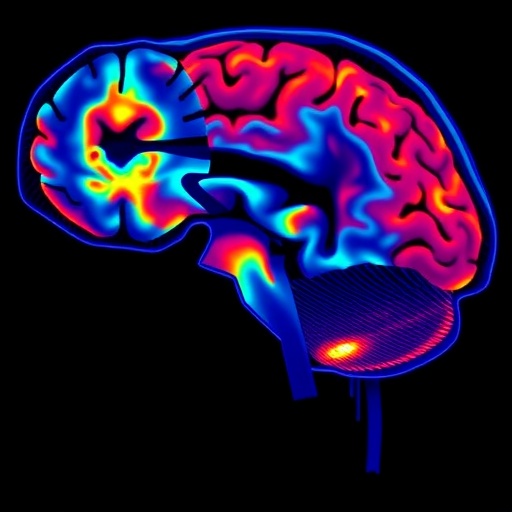
Neuromelanin, a dark pigment found within specific populations of neurons in the substantia nigra—a brain region critically affected in PD—has emerged as a focal point for imaging innovations. Unlike conventional neuroimaging that targets gross anatomical changes or nonspecific markers of neurodegeneration, neuromelanin imaging directly visualizes this pigment’s distribution and density, reflecting the health and presence of dopaminergic neurons. As these neurons degenerate in Parkinson’s disease, neuromelanin content diminishes, providing a direct, quantifiable correlate of neuronal loss that can be detected by magnetic resonance imaging.
The study under scrutiny meticulously compared two cutting-edge MRI modalities: neuromelanin-sensitive imaging and free water imaging. Free water imaging quantifies the presence of extracellular water, which increases in brain tissue degeneration and neuroinflammation. While this method has shown promise in detecting microstructural changes associated with PD, it lacks the biochemical specificity offered by neuromelanin imaging. By enrolling a cohort of patients at the earliest clinically diagnosable stage of Parkinson’s, alongside matched healthy controls, the researchers executed a rigorous protocol designed to evaluate and contrast the diagnostic power of these techniques.
Statistically robust analyses revealed that neuromelanin imaging consistently exhibited higher sensitivity and specificity in identifying subtle neurodegenerative changes within the substantia nigra compared to free water imaging. Particularly compelling was the ability of neuromelanin imaging to detect these alterations before significant motor symptoms emerged, highlighting its potential as a preclinical biomarker. Furthermore, neuromelanin imaging showed strong correlations with clinical rating scales of motor impairment and disease severity, underscoring its clinical relevance and translational applicability.
The implications of these findings extend far beyond diagnostic enhancement. By accurately mapping neuromelanin loss, clinicians and researchers can gain deeper insight into disease trajectory and heterogeneity, which are crucial for tailoring individualized treatment plans. Additionally, neuromelanin imaging could serve as a valuable surrogate endpoint in clinical trials, accelerating the development of therapeutic agents by providing objective and quantifiable measures of neuroprotection or neurorestoration.
On a technical level, the neuromelanin imaging technique leverages specialized MRI sequences sensitive to the paramagnetic properties of neuromelanin-bound metals, such as iron. This approach captures contrast differences without the need for exogenous contrast agents, minimizing patient risk. Advances in scanner technology and sequence optimization have enhanced resolution and signal-to-noise ratios, overcoming previous barriers that limited neuromelanin imaging feasibility in clinical settings.
Nevertheless, challenges remain in standardizing neuromelanin imaging protocols across different MRI platforms and centers, ensuring reproducibility and comparability essential for widespread adoption. The integration of artificial intelligence and machine learning for automated image analysis holds promise in accelerating data interpretation and reducing observer bias. Moreover, multidisciplinary collaborations involving neurologists, radiologists, and bioengineers are imperative to translate these innovations from bench to bedside effectively.
This study also prompts a re-evaluation of existing neurodegenerative disease frameworks by introducing biochemical specificity to in vivo imaging biomarkers. While free water imaging offers valuable insights into neuroinflammatory processes and tissue microenvironment changes, its inability to definitively pinpoint dopaminergic neuron loss limits its diagnostic clinch. Neuromelanin imaging, by contrast, provides a window into the actual neurodegenerative substrate—loss of pigment-rich neurons—that directly underpin Parkinsonian pathology.
Looking forward, combining neuromelanin imaging with other emerging modalities, such as alpha-synuclein PET imaging or ultrahigh-field MRI, could yield multimodal biomarkers that capture diverse facets of Parkinson’s disease pathology. Such integrative approaches may unlock sophisticated diagnostic algorithms capable of distinguishing Parkinson’s disease from other parkinsonian syndromes with overlapping clinical profiles, a critical step towards precision neurology.
The broader neuroscience community is already lauding this study as a tour de force proof-of-concept that could inspire biomarker exploration for other neurodegenerative disorders where pathognomonic pigments or compounds exist. In addition to Parkinson’s, conditions such as Alzheimer’s disease—where tau and beta-amyloid imaging have revolutionized diagnosis—could benefit from analogous pigment-specific imaging strategies, potentially heralding a new era of disease-specific molecular neuroimaging.
From a clinical perspective, earlier and more accurate diagnosis enabled by neuromelanin imaging could mitigate the enormous healthcare burden posed by PD through timely intervention, patient education, and support services. Moreover, this technique’s non-invasive nature and reliance on widely available MRI infrastructure make it an accessible tool for routine neurological practice, potentially democratizing advanced diagnostics beyond specialized centers.
In sum, the research presented by Roh et al. marks a paradigm shift in Parkinson’s disease detection, with neuromelanin imaging setting a new benchmark for early, reliable, and pathophysiologically relevant diagnosis. As these findings permeate clinical workflows and inspire further technological refinements, patients stand to benefit from earlier therapeutic windows, optimized management strategies, and ultimately, improved outcomes. The quest for conquering Parkinson’s disease just gained a powerful ally in neuromelanin-sensitive MRI.
The future promises an exciting intersection of neuroimaging science, clinical neurology, and therapeutic innovation centered on this novel approach. Continued validation studies, enhanced imaging protocols, and integration with computational analytics will solidify neuromelanin imaging as a cornerstone of Parkinson’s disease diagnostics. With neurologists armed with sharper vision into the brain’s darkest pigments, the hope for intercepting neurodegeneration at its origin moves ever closer to reality.
Subject of Research: Early diagnosis of Parkinson’s disease using advanced MRI techniques
Article Title: Neuromelanin imaging outperforms free water imaging in diagnosing early Parkinson’s disease: a comparative MRI study
Article References:
Roh, Y.H., Youn, J., Kim, SY. et al. Neuromelanin imaging outperforms free water imaging in diagnosing early Parkinson’s disease: a comparative MRI study. npj Parkinsons Dis. (2026). https://doi.org/10.1038/s41531-026-01286-y
Image Credits: AI Generated
Tags: advanced MRI techniques for Parkinson’sclinical implications of neuromelanin MRIearly detection of Parkinson’s diseaseearly-stage Parkinson’s neuroimagingfree water imaging limitations in Parkinson’simproving Parkinson’s diagnostic accuracyneuromelanin imaging vs free water imagingneuromelanin MRI in early Parkinson’s diagnosisnovel neuroimaging biomarkers for Parkinson’sParkinson’s disease neurodegeneration biomarkerssubstantia nigra neuromelanin imagingtherapeutic intervention timing in Parkinson’s
