Neonatal encephalopathy (NE) remains one of the most pressing challenges in pediatric neurology, given its profound impact on infant survival and long-term neurodevelopmental outcomes worldwide. At its core, NE represents a syndrome of disturbed neurological function in newborns, predominantly caused by hypoxic-ischemic events during the perinatal period. Despite advances in medical care, it continues to be the primary driver of lifelong disabilities including cerebral palsy, cognitive impairment, and deficits in behavior and executive functioning. The complexity of the condition stems not only from its multifactorial etiology but also from the evolving nature of clinical presentations, complicating early diagnosis and prognostication efforts.
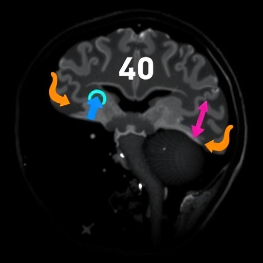
In the quest to unravel the intricate brain injuries underlying neonatal encephalopathy, magnetic resonance imaging (MRI) has emerged as the definitive tool. Unlike other imaging modalities, MRI offers unparalleled soft tissue contrast and exquisite anatomical detail, essential for delineating the extent and pattern of cerebral injury. Among MRI techniques, diffusion-weighted imaging (DWI) has revolutionized early detection capabilities, as it sensitively captures the early cytotoxic edema that typifies hypoxic-ischemic injury. Through the measurement of water molecule displacement at a microscopic scale, DWI allows clinicians to detect brain areas undergoing acute stress within hours of insult, dramatically influencing therapeutic decisions.
Complementing DWI, proton magnetic resonance spectroscopy (^1H-MRS) provides a metabolic window into the infant brain. This technique measures the concentration of various brain metabolites, with the lactate to N-acetylaspartate (Lac/NAA) peak area ratio serving as a particularly reliable biomarker. Elevated lactate reflects anaerobic metabolism induced by hypoxia, while reductions in NAA signify neuronal loss or dysfunction. The combined assessment from the basal ganglia and thalamus regions affords a robust biochemical signature that correlates strongly with two-year neurodevelopmental outcomes. Such molecular insights extend beyond anatomical imaging, offering predictive power that guides clinical management and family counseling.
The development of multimodal MRI scoring systems marks a significant leap forward in the prognostic evaluation of NE. By integrating data from conventional MRI, DWI, and MRS, these composite scales achieve superior correlation with neurodevelopmental milestones, facilitating individualized prognosis. The synergy achieved in combining structural and metabolic information underscores the necessity of comprehensive imaging approaches. Each modality captures different facets of the brain’s injury landscape – from gross anatomical disruptions to subtle biochemical alterations – rendering a holistic perspective that no solitary method can provide.
Beyond the traditional realms of MRI and spectroscopy, advances in neuroimaging continue to push the boundaries of understanding neonatal brain injury at a microstructural and functional level. Diffusion tensor imaging (DTI) dissects white matter integrity by tracking anisotropic water diffusion along axonal tracts, shedding light on connectivity disruptions invisible on standard MRI. Similarly, arterial spin labeling (ASL) non-invasively measures cerebral perfusion by magnetically tagging blood water molecules, allowing assessment of regional blood flow changes in compromised brain regions. Functional MRI, harnessing blood oxygen level-dependent (BOLD) contrast, offers dynamic insights into brain activity and network connectivity, potentially unmasking functional deficits that arise from injury.
Standardization emerges as a crucial theme in advancing MRI biomarkers from research tools to clinical mainstays. Harmonizing acquisition protocols and post-processing pipelines ensures reproducibility and comparability across centers and studies, a prerequisite for reliable biomarker validation. This standardization not only accelerates the translation of neuroimaging findings into routine clinical care but also enhances the power of neuroprotection trials. By providing early surrogate endpoints that closely predict long-term outcomes, MRI biomarkers enable trials with smaller sample sizes and faster timelines, hastening the advent of novel therapeutics.
The interplay between MRI and ^1H-MRS represents a paradigm shift in neonatal encephalopathy care. Where once prognosis relied heavily on clinical scoring and physiological parameters, the integration of imaging biomarkers provides objective, quantifiable metrics of brain injury severity. This convergence informs critical decision-making, from therapeutic hypothermia eligibility to anticipatory guidance for families regarding developmental expectations. Furthermore, the evolving consensus underscores the pressing need to incorporate imaging into standard neurocritical care pathways, ensuring timely and targeted interventions.
Therapeutic hypothermia, while revolutionary in reducing mortality and improving outcomes, remains insufficient for a substantial subset of infants with NE. Many survivors still bear significant neurodevelopmental disabilities, highlighting the urgent imperative to refine prognostic tools and to develop adjunctive neuroprotective strategies. Advanced neuroimaging modalities offer hope not only for enhanced prediction but also for monitoring therapeutic efficacy, enabling real-time adjustments and personalized treatment paradigms.
As research progresses, the role of MRI biomarkers in clinical trials extends beyond outcome prediction to serve as surrogate endpoints. Their sensitivity to subtle brain changes offers critical advantages in evaluating new therapeutic agents or protocols. This capacity to detect early neuroprotective effects or identify emerging injury trends can dramatically reduce the duration and cost of trials, fostering rapid innovation in NE management. Moreover, such biomarkers lay the groundwork for precision medicine approaches, stratifying patients based on injury profiles and likely trajectories.
In addition to technical advances, interdisciplinary collaboration remains pivotal in translating MRI and spectroscopy insights into improved patient care. Radiologists, neonatologists, neurologists, and researchers must synergize efforts to refine imaging protocols, interpret complex data, and validate findings against neurodevelopmental outcomes. Training programs in neonatal neuroimaging interpretation and the deployment of centralized image repositories could further enhance expertise dissemination and benchmarking.
The promise of advanced neuroimaging extends beyond immediate neonatal care to influence long-term surveillance and intervention strategies. By charting the evolution of brain injury and recovery, serial MRI assessments can guide rehabilitation efforts, identify windows of neuroplasticity, and inform educational planning. This lifelong perspective emphasizes the foundational role of precise early imaging in optimizing developmental trajectories and quality of life for affected children.
Future opportunities abound as MRI technology continues to evolve. Ultrahigh-field MRI scanners, quantitative susceptibility mapping, and machine learning-assisted image analysis represent frontiers that could deepen insight into neonatal brain injury pathophysiology. Machine learning algorithms, in particular, hold potential for automating image interpretation, standardizing scoring, and integrating multimodal data into predictive models with unprecedented accuracy.
In conclusion, magnetic resonance imaging and spectroscopy have redefined the landscape of neonatal encephalopathy diagnosis and prognosis. Their integration provides a powerful, multifaceted understanding of brain injury patterns, biochemical changes, and functional disruptions. As consensus aligns on standardized protocols and clinical applicability, these imaging modalities are set to become indispensable tools in neonatology. Their influence extends from bedside decision-making to accelerating neuroprotection clinical trials and fostering precision pediatric neurology – a promising horizon for the care of the most vulnerable patients.
Subject of Research: Neonatal encephalopathy; neuroimaging biomarkers; prognostication and outcomes; magnetic resonance imaging and spectroscopy in neonatal brain injury.
Article Title: Magnetic resonance imaging and spectroscopy in neonatal encephalopathy: current consensus position and future opportunities.
Article References:
Laptook, A., Garvey, A.A., Adams, C. et al. Magnetic resonance imaging and spectroscopy in neonatal encephalopathy: current consensus position and future opportunities. Pediatr Res (2025). https://doi.org/10.1038/s41390-025-04448-5
Image Credits: AI Generated
DOI: https://doi.org/10.1038/s41390-025-04448-5
Tags: advances in MRI technologycerebral palsy risk factorscognitive impairment in infantsdiffusion-weighted imaging applicationsearly detection of brain injuryhypoxic-ischemic brain injurylong-term neurodevelopmental outcomesMRI and spectroscopy techniquesneonatal brain injury diagnosisneonatal encephalopathypediatric neurology challengesprognostication in neonatal care


