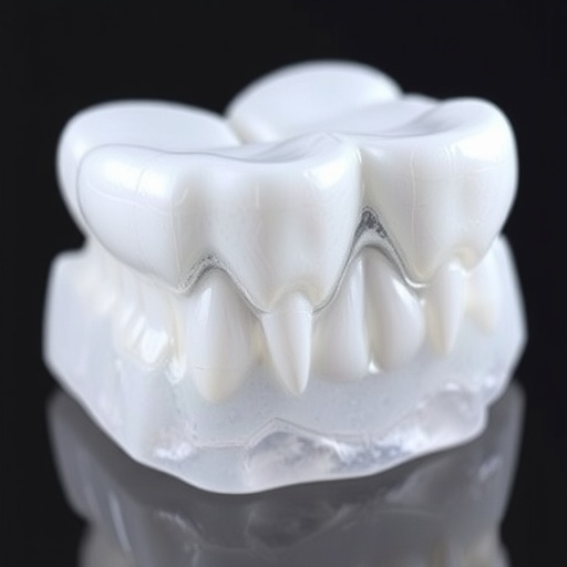
In a groundbreaking study published in BioMedical Engineering OnLine, researchers have unveiled remarkable advances in dental restorative technology, demonstrating that nano-hydroxyapatite (nano-HA) fillers significantly enhance masticatory function and modulate inflammatory markers in patients with periapical inflammation. This pioneering research addresses long-standing challenges faced in endodontic therapy, offering a promising alternative to conventional root canal filling materials by harnessing the unique properties of nano-scale hydroxyapatite particles.
Periapical inflammation represents a frequent and painful dental condition, often resulting from bacterial infection within the root canal system. Traditional treatment strategies typically involve mechanical debridement and filling of the canal with inert materials; however, these approaches do not actively promote tissue regeneration or control ongoing inflammation efficiently. In this context, the study led by Xie et al. evaluates how the incorporation of nano-HA as a filling material impacts both functional recovery and biochemical inflammatory responses in gingival sulcular fluid, which is critical to oral healing and long-term clinical success.
The research enrolled 98 patients diagnosed with periapical inflammation, who were randomly assigned to either a control group receiving standard root canal therapy or an experimental group treated with nano-HA filling. This randomized controlled design ensured an unbiased comparison between the innovative nano-HA technique and conventional approaches. Measurements were meticulously taken before treatment, one week after intervention, and at a three-month follow-up, focusing on functional parameters like bite force and masticatory efficiency, alongside inflammatory cytokine levels such as interleukin-1β (IL-1β) and tumor necrosis factor-α (TNF-α).
One of the most striking findings of the study is the superior improvement in masticatory performance observed in the nano-HA group. Patients treated with nano-HA experienced marked increases in bite force and chewing efficiency compared to the control cohort, highlighting the potential of nano-HA to restore oral function more effectively. This enhancement likely stems from the biomimetic nature of hydroxyapatite, a calcium phosphate mineral that constitutes the primary inorganic component of human bone and dental enamel, making it inherently compatible with tooth tissue.
Furthermore, the study reveals profound biochemical benefits associated with nano-HA filling. Levels of pro-inflammatory cytokines IL-1β and TNF-α in gingival sulcular fluid demonstrated a continuous decline in the experimental group, indicating a robust anti-inflammatory effect. These cytokines are key mediators in periapical inflammation, driving tissue destruction and pain. The ability of nano-HA to actively suppress their expression suggests it not only serves as a filler but also modulates the local immune response to foster a more conducive environment for healing.
The clinical outcomes also extended to enhanced filling effects and accelerated healing rates. Radiographic and clinical assessments confirmed that the nano-HA treated group exhibited better sealing of the root canal system with fewer signs of recurrent infection or inflammation. This can be attributed to the nano-scale dimensions of hydroxyapatite particles, which enable tighter packing and superior interface bonding with dentin. This nano-structural characteristic may prevent microleakage, a common cause of root canal therapy failure.
Importantly, the study identified a negative correlation between the application of nano-HA fillers and inflammatory marker levels at both one week and three months post-treatment, underlining a sustained therapeutic impact beyond the immediate post-operative period. This sustained reduction in inflammation could translate into long-term preservation of periodontal health, which is crucial in preventing tooth loss and maintaining oral quality of life.
The utilization of nano-HA in dental restoration is anchored in its excellent biocompatibility and osteoconductivity, properties extensively documented in bone tissue engineering literature. The current investigation extends these advantages into the delicate environment of the dental pulp and periodontal ligament spaces, demonstrating that nano-HA not only supports structural restoration but also biological homeostasis. This dual function elevates it beyond conventional inert filling materials.
Moreover, the influence of nano-HA on masticatory muscle function could be mediated through improved proprioception and biomechanical stability of the restored tooth. As bite force and chewing efficiency are vital indicators of oral health, their enhancement points to significant quality-of-life improvements for patients suffering from periapical pathology. This multidimensional benefit underscores the transformative potential of nano-HA in clinical endodontics.
While the study holds immense promise, it also invites further exploration into the mechanisms underlying the anti-inflammatory properties of nano-HA. Prior hypotheses suggest that hydroxyapatite nanoparticles may interact with local immune cells, modulating signaling pathways that govern cytokine production. Deciphering these pathways could pave the way for customized nanomaterial therapies tailored to individual inflammatory profiles.
Additionally, long-term clinical trials are necessary to validate these promising short and intermediate-term findings. Assessing factors such as longevity of the filling, resistance to bacterial colonization, and structural integrity under functional load will solidify the case for widespread adoption of nano-HA in dental practices worldwide. The excitement surrounding these results calls for integration of nanotechnology with traditional dentistry to revolutionize patient outcomes.
The present study stands as a testament to the rapidly evolving interface between materials science and biomedical engineering. Through precise design and application of nano-scale biomaterials, clinicians are equipped with novel tools that not only restore structure but also guide biological healing processes. Such advances resonate beyond dentistry, potentially informing regenerative strategies in broader musculoskeletal contexts.
In conclusion, the compelling evidence presented by Xie and colleagues advocates for the clinical promotion of nano-HA restorative materials in the management of periapical inflammation. Their work not only demonstrates enhanced masticatory function and inflammatory control but also sets a precedent for future material innovations that prioritize biological functionality alongside mechanical performance. As the dental community seeks to improve patient care through technology, nano-hydroxyapatite emerges as a frontrunner in the quest for superior endodontic solutions.
Subject of Research: The effect of nano-hydroxyapatite filling on masticatory function and inflammatory factor levels in gingival sulcular fluid in patients with periapical inflammation.
Article Title: Effect of nano-hydroxyapatite filling on masticatory function and gingival sulcular fluid inflammatory factor levels in periapical inflammation.
Article References:
Xie, H., Tan, A., Su, Z. et al. Effect of nano-hydroxyapatite filling on masticatory function and gingival sulcular fluid inflammatory factor levels in periapical inflammation. BioMed Eng OnLine 24, 63 (2025). https://doi.org/10.1186/s12938-025-01374-9
Image Credits: AI Generated
DOI: https://doi.org/10.1186/s12938-025-01374-9
Tags: alternative dental therapiesclinical success in dental treatmentsdental restorative technology advancementsendodontic therapy innovationsgingival sulcular fluid analysisinflammatory marker modulationmasticatory function enhancementnano-hydroxyapatite in dentistryoral healing and regenerationperiapical inflammation treatmentrandomized controlled trial in dentistryroot canal filling materials


