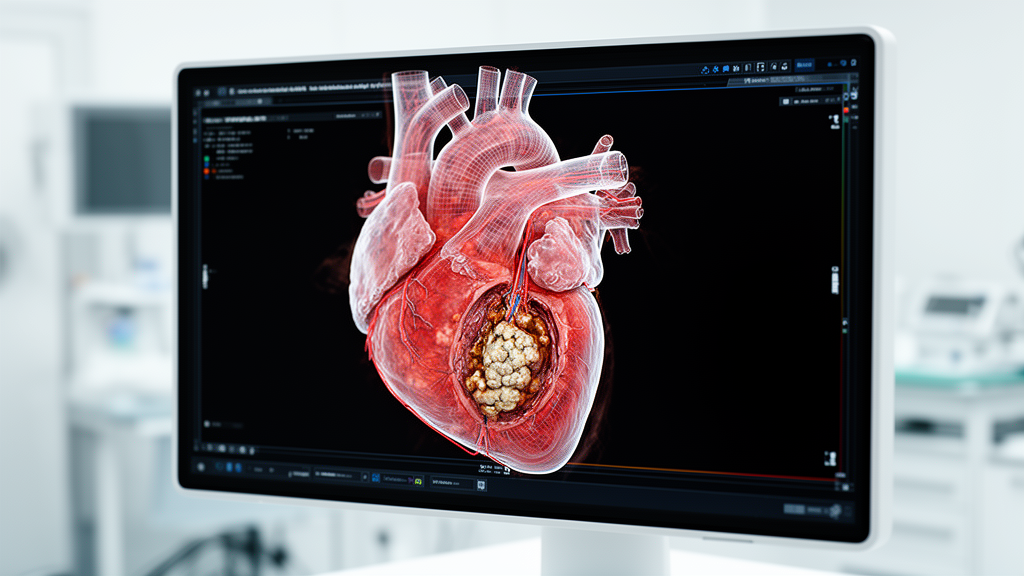
A narrative review published in Frontiers in Cardiovascular Medicine outlines a structured, phenotype-first multimodality imaging framework designed to resolve diagnostic challenges in discordant low-gradient aortic stenosis. Severe aortic stenosis has traditionally been identified when an aortic valve area of 1.0 square centimetre or less coincides with a mean transvalvular gradient of at least 40 millimetres of mercury and a peak velocity of at least 4.0 metres per second. However, clinical registries indicate that up to 40 percent of patients evaluated for aortic stenosis in selected echocardiographic cohorts exhibit a discordant low-gradient pattern, combining a calculated aortic valve area of 1.0 square centimetre or less with a mean gradient below 40 millimetres of mercury and a peak velocity under 4.0 metres per second. This profile creates substantial clinical ambiguity because determining whether valve obstruction is genuinely severe directly dictates whether an individual patient should be assigned to clinical surveillance, undergo advanced imaging adjudication, or proceed to surgical or transcatheter aortic valve replacement.
Misclassifying aortic stenosis carries considerable clinical risk in both directions. Underestimating true-severe obstruction may lead clinicians to defer valve intervention until irreversible myocardial decompensation has developed, while erroneously categorising pseudo-severe or moderate disease as severe exposes individuals to unnecessary procedural work-up and replacement hazards. Review authors Yuan Li, Xue An, and Yujian Liu emphasise that existing guidelines from major cardiovascular societies already provide essential diagnostic pathways, such as verifying echocardiographic measurement quality, classifying cases by left ventricular ejection fraction and stroke volume index, employing dobutamine stress echocardiography in classical low-flow states, and performing computed tomography calcium scoring in preserved ejection fraction presentations. Nevertheless, persistent diagnostic uncertainty frequently arises when haemodynamic measurements, anatomical valve calcification, clinical symptoms, and myocardial markers provide conflicting signals. The authors propose that the remaining clinical challenge is primarily operational, requiring consistent documentation of transvalvular flow rates, systematic reporting of technical study adequacy, phenotype-specific weighting of diagnostic modalities, and clear communication of residual diagnostic uncertainty.
The physical basis of low-gradient discordance stems directly from the flow dependency of transvalvular pressure gradients. In accordance with the simplified Bernoulli formulation, the pressure gradient across a stenotic valve varies with the square of flow velocity. Consequently, when forward stroke volume or instantaneous transvalvular flow rate declines, the measured transvalvular gradient can drop well below the standard 40 millimetres of mercury threshold despite the presence of rigid, severe anatomical obstruction. In addition, calculated valve areas derived via the continuity equation remain vulnerable to measurement error because left ventricular outflow tract diameter is squared in the continuity calculation, meaning small linear measurement discrepancies can inappropriately reclassify a patient across the severe disease threshold. Technical confounders such as Doppler beam misalignment, incomplete acoustic window interrogation, incorrect velocity time integral tracing, irregular cardiac rhythms, uncontrolled hypertension, and concomitant valvular disease can all mimic genuine pathophysiological discordance.
To establish diagnostic clarity, the framework categorises patients into distinct haemodynamic phenotypes before advanced diagnostic tests are ordered or interpreted. Classical low-flow, low-gradient aortic stenosis is defined by an aortic valve area of 1.0 square centimetre or less, a mean gradient below 40 millimetres of mercury, a left ventricular ejection fraction below 50 percent, and a stroke volume index below 35 millilitres per square metre. The review highlights that while categorical cut-offs provide helpful initial grouping, stroke volume index represents a continuous haemodynamic parameter that must be interpreted in conjunction with sex, body size, ejection time, and transvalvular flow rate. In this classical phenotype, the primary clinical inquiry focuses on distinguishing true-severe stenosis from pseudo-severe valve narrowing caused by impaired left ventricular contractility that cannot generate sufficient force to open the leaflets fully.
A second major phenotype, paradoxical low-flow, low-gradient aortic stenosis, occurs when patients maintain a preserved left ventricular ejection fraction of 50 percent or higher despite experiencing a reduced forward stroke volume index below 35 millilitres per square metre. This physiology frequently coincides with pronounced concentric ventricular remodelling, small ventricular cavity dimensions, marked diastolic dysfunction, elevated global afterload, atrial fibrillation, and reduced longitudinal systolic strain despite a preserved volumetric ejection fraction. The therapeutic benefit of aortic valve replacement remains less clearly established in this population because the low transvalvular gradient may reflect systemic vascular changes and extracardiac contributors to low forward flow rather than severe intrinsic leaflet obstruction. In addition, cardiac amyloidosis represents a vital diagnostic red flag in paradoxical low-flow states, requiring targeted monoclonal protein screening and bone scintigraphy when disproportionate wall thickness, low electrocardiographic voltage, or apical-sparing strain patterns appear.
Normal-flow low-gradient aortic stenosis represents the most contested subgroup, characterised by an aortic valve area of 1.0 square centimetre or less, a mean gradient below 40 millimetres of mercury, an ejection fraction of at least 50 percent, and a stroke volume index of 35 millilitres per square metre or higher. The review authors note that this subgroup brings together a heterogeneous mixture of genuine severe obstruction, moderate disease misclassified by outflow tract measurement errors, normal physiology in individuals with small body surface area, and states with reduced instantaneous transvalvular flow despite preserved stroke volume. Cohort studies cited in the review, including investigations from the SEAS trial and retrospective clinical cohorts, demonstrate that patients with normal-flow low-gradient disease frequently experience clinical trajectories comparable to moderate aortic stenosis. Consequently, the proposed framework recommends treating normal-flow low-gradient cases as a diagnostic audit category, recalculating transvalvular flow rate using proposed thresholds around 200 to 210 millilitres per second, and avoiding automatic referral for intervention unless structural and clinical parameters clearly converge.
A critical initial phase of the framework involves rigorous echocardiographic quality control prior to testing escalation. Clinicians are directed to verify that the left ventricular outflow tract diameter is measured precisely at the aortic annulus or immediate cusp insertion plane, as measuring five to ten millimetres apically into the muscular outflow tract systematically underestimates stroke volume and valve area. Interrogating continuous-wave Doppler signals across multiple acoustic windows, specifically including the right parasternal window, reduces gradient underestimation. When two-dimensional geometry remains uncertain, calculating hybrid valve areas using cross-sectional outflow tract dimensions from transoesophageal echocardiography, computed tomography, or cardiovascular magnetic resonance can clarify orifice dimensions. Furthermore, the review advises that direct anatomical planimetry derived from cross-sectional imaging must be interpreted against an anatomical reference cut-off of approximately 1.2 square centimetres rather than the functional 1.0 square centimetre continuity threshold.
For physiological severity adjudication in classical low-flow, low-gradient disease, dobutamine stress echocardiography remains the primary functional evaluation tool. Flow reserve is defined by a 20 percent or greater increase in stroke volume during dobutamine infusion. The review notes data from the TOPAS study showing that traditional criteria—such as relying on a peak-stress mean gradient of at least 40 millimetres of mercury or peak valve area of 1.0 square centimetre or less—correctly classified stenosis severity in only 48 percent and 60 percent of patients, respectively. In contrast, calculating projected aortic valve area at a standardised flow rate achieved a 70 percent correct classification rate, demonstrating 86 percent sensitivity and 47 percent specificity, with a projected valve area of 1.0 square centimetre or less independently predicting mortality in conservatively managed individuals with a hazard ratio of 3.65. The authors highlight that the absence of flow reserve should not preclude intervention, as contemporary registry analyses indicate clinical benefit from transcatheter replacement even without demonstrable contractile reserve.
Non-contrast, electrocardiographically gated computed tomography aortic valve calcium scoring provides an anatomical assessment that remains completely independent of loading conditions and transvalvular flow rates. In a multicentre validation study of 918 patients across eight centres, calcium scoring exhibited high diagnostic discrimination for severe stenosis, yielding area under the curve values of 0.92 in women and 0.89 in men, with optimal thresholds identified at 1,377 Agatston units for women and 2,062 Agatston units for men. The proposed framework applies likelihood tiers adapted from professional guidelines, defining calcific severity as highly likely above 3,000 Agatston units in men and 1,600 in women, and unlikely below 1,600 in men and 800 in women. The authors caution that calcium scoring can underestimate disease severity in younger patients, individuals with bicuspid aortic valves, and women, who tend to develop a higher proportion of fibrotic rather than calcific leaflet thickening, making contrast-enhanced computed tomography fibrocalcific assessment a valuable supplementary tool.
A central tenet of the framework is the strict operational separation of valvular severity adjudication from myocardial risk refinement. Advanced markers of myocardial damage—including replacement fibrosis detected by late gadolinium enhancement on magnetic resonance, impaired global longitudinal strain, expanded extracellular volume fraction, and anatomical staging of downstream cardiac damage—provide valuable prognostic stratification but should not serve as standalone triggers for valve replacement. Observational data demonstrate that myocardial replacement scar independently predicts mortality, with each additional one percent of scar volume associated with an 11 percent increase in all-cause mortality hazard, and long-term follow-up showing a 3.5-year reduction in median survival. However, findings from the randomised EVOLVED trial, which evaluated early intervention guided by late gadolinium enhancement, yielded neutral results, cautioning against using fibrosis alone to mandate procedural intervention in the absence of verified severe hemodynamic obstruction.
The review culminates in a structured Heart Team communication template designed to replace ambiguous binary reporting with transparent likelihood tiers. Rather than forcing a definitive diagnosis when imaging findings diverge, the framework standardises three distinct clinical outputs: true-severe aortic stenosis likely, non-severe or pseudo-severe stenosis likely, or unresolved discordance. When technical limitations, indeterminate calcium scores within sex-specific grey zones, or conflicting flow dynamics prevent definitive classification, the clinical report explicitly documents the specific sources of uncertainty, technical limitations, and the precise diagnostic test that could alter patient management. The authors conclude that while the framework structures multidisciplinary Heart Team deliberations and reduces misclassification, prospective multicentre validation remains necessary before the pathway can be deployed as an autonomous treatment algorithm.
Subject of Research: Multimodality imaging framework for evaluating discordant low-gradient aortic stenosis phenotypes
Article Title: Discordant low-gradient aortic stenosis: a phenotype-first, likelihood-tiered multimodality imaging framework
Article References: Li, Y., An, X., & Liu, Y. (2026). Discordant low-gradient aortic stenosis: a phenotype-first, likelihood-tiered multimodality imaging framework. Frontiers in Cardiovascular Medicine, 13, Article 1901893. https://doi.org/10.3389/fcvm.2026.1901893
Image Credits: AI Generated
DOI: 10.3389/fcvm.2026.1901893
Keywords: aortic stenosis, discordant low-gradient aortic stenosis, multimodality imaging, dobutamine stress echocardiography, CT aortic valve calcium scoring, echocardiography, cardiovascular magnetic resonance, stroke volume index, transvalvular flow rate, projected aortic valve area, late gadolinium enhancement, global longitudinal strain, cardiac damage staging, heart team, cardiac amyloidosis, hemodynamics
News Source: Ophelia Keating. (November 14, 2026). Multimodality Imaging Framework Refines Evaluation of Discordant Low Gradient Aortic Stenosis. Scienmag.