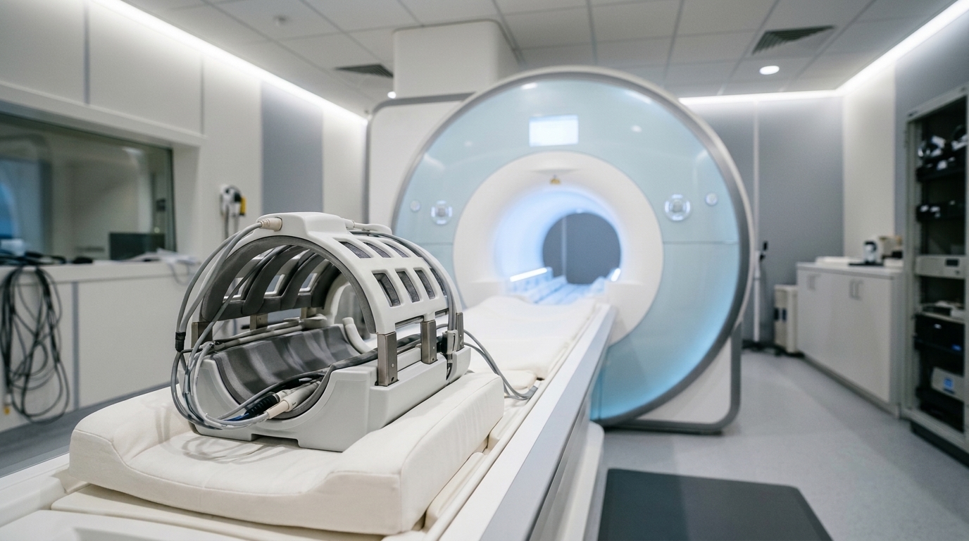
A cross-sectional neuroimaging investigation utilizing multidimensional magnetic resonance imaging has revealed that normative human brain aging is characterized by a coordinated redistribution of microstructural features across cellular-scale dimensions rather than the emergence of entirely new tissue components. By evaluating forty-six cognitively unimpaired adults spanning an age range from twenty to seventy-seven years, researchers demonstrated that age-dependent structural reorganization unfolds simultaneously across cellular shape, microscopic size, barrier restriction, and local chemical environment. The multidimensional imaging framework departs fundamentally from traditional neuroimaging paradigms that assign a single averaged scalar value to each imaging voxel, instead resolving intra-voxel distributions of diffusion and relaxation properties without imposing rigid geometric assumptions. The resulting empirical phenotypes mapped systematic structural alterations across cortical gray matter, subcortical nuclei, and major white matter tracts throughout the adult lifespan. Specifically, the data demonstrated widespread shifts from small length-scale environments toward intermediate and larger length scales in gray matter, high-to-medium anisotropy transitions across major white matter pathways, and prominent accumulations of fast transverse relaxation phenotypes within deep subcortical structures.
Understanding the precise biological progression of brain aging requires resolving structural alterations across vastly disparate spatial scales, from gross anatomical degeneration to microscopic cytoarchitectural remodeling. Concurrently, white matter pathways undergo macroscopic deterioration characterized by loss of tract coherence, reduced myelination, and cumulative pathway disconnection. However, postmortem histological analyses reveal that these macroscopic manifestations represent relatively late or cumulative endpoints of subtle microscopic processes that begin decades earlier. Early cellular-scale aging involves progressive dendritic spine regression, synaptic pruning, reactive glial remodeling, and gradual enlargement of the interstitial extracellular space. Although standard diffusion tensor imaging captures bulk alterations such as elevated mean diffusivity and decreased fractional anisotropy, and biophysical compartment models like NODDI or SANDI attempt to estimate neurite density, these established techniques depend heavily on predetermined geometric assumptions that falter in complex, isotropic, or weakly anisotropic brain regions.
To overcome the intrinsic physical and mathematical limitations of scalar imaging metrics and biophysical compartment models, researchers implemented an advanced multidimensional magnetic resonance imaging framework that jointly measures diffusion tensors and relaxation rates. This innovative imaging strategy integrates tensor-valued diffusion encoding with longitudinal and transverse relaxometry, allowing water molecules to probe distinct microenvironments characterized by specific boundaries, membrane geometries, and macromolecular interactions. Rather than forcing continuous biological measurements into predefined compartments such as idealized cylinders or spheres, multidimensional magnetic resonance imaging reconstructs joint continuous parameter spectra directly from acquired radiofrequency signals. A key technical feature of this implementation is the incorporation of frequency-dependent diffusion measurements, which evaluate how water displacement varies across specific gradient oscillation frequencies. Because time-dependent diffusion directly reflects physical restrictions imposed by cellular membranes and subcellular organelles, measuring the diffusion frequency spectrum provides explicit sensitivity to structural barriers at the micrometer scale. This noninvasive approach disentangles coexisting tissue microenvironments that previously appeared blurred or indistinguishable within conventional imaging voxels.
The data acquisition protocol was executed on a clinical three-tesla MAGNETOM Prisma scanner equipped with a thirty-two-channel receive head array at the National Institute on Drug Abuse. Multidimensional diffusion-relaxometry scans were acquired with a single-shot spin-echo echo-planar imaging sequence incorporating numerically optimized, asymmetric gradient waveforms that generated linear, spherical, and planar diffusion-encoding b-tensors. Longitudinal and transverse relaxation sensitivities were systematically established by combining six distinct repetition times ranging from 0.62 to 7.6 seconds with four distinct echo times spanning 40 to 150 milliseconds, yielding a total of 139 unique multidimensional diffusion-relaxation encoding combinations collected over approximately forty minutes per participant. Anatomical images were simultaneously acquired at one-millimeter isotropic resolution using a fat-suppressed T1-weighted MPRAGE sequence to guide spatial co-registration, skull stripping, and precise region-of-interest segmentation across twenty-eight predefined cortical, subcortical, and white matter structures.
Following raw data acquisition, comprehensive computational preprocessing was performed to eliminate spatial distortions, correct for head motion, and reconstruct continuous parameter spectra. Diffusion volumes were denoised using Marchenko-Pastur principal component analysis before undergoing affine and quadratic transformations in the TORTOISE software package to correct for eddy currents and subject displacement, which remained remarkably low across the entire cohort between 0.24 and 0.85 millimeters. Susceptibility-induced geometric distortions were corrected through the DR-BUDDI algorithm utilizing reversed phase-encoded blips alongside synthetic anatomical targets. Preprocessed signals were subsequently inverted via a Monte Carlo framework to reconstruct three-dimensional joint distributions of isotropic diffusivity, squared microscopic diffusion anisotropy, and transverse relaxation rates. Researchers subsequently established ten empirical microstructural phenotypes by partitioning continuous parameter dimensions into heuristic subdomains: low, intermediate, and high isotropic diffusivity capturing small, intermediate, and large length scales; low, intermediate, and high microscopic anisotropy reflecting spherical, eccentric, and highly directional cellular geometries; and low versus high transverse relaxation rates distinguishing free tissue water from macromolecule-associated or iron-influenced environments.
Analysis of white matter tracts revealed a coherent, highly constrained pattern of microstructural reorganization across the adult lifespan. In compact commissural tracts such as the genu of the corpus callosum, younger adults exhibited a prominent, highly restricted microstructural phenotype characterized by low isotropic diffusivity, high microscopic diffusion anisotropy, and fast transverse relaxation. As age advanced, this dominant high-anisotropy phenotype demonstrated a significant, systematic transition toward intermediate microscopic anisotropy accompanied by reductions in the fast relaxation component, without the emergence of entirely novel spectral components. Group-difference maps confirmed that aging in white matter pathways involves a direct signal migration from highly directional, tightly packed axonal environments toward less anisotropic, moderately dispersed regimes alongside unconstrained, high-diffusivity subdomains. Furthermore, significant negative linear associations with age were observed for high-anisotropy, fast-relaxing signal fractions within the anterior corona radiata, superior fronto-occipital fasciculus, and genu of the corpus callosum. In the context of known neurobiology, these coordinated white matter transitions reflect progressive age-related myelin degradation, loosening of tight axonal packing, increased orientational fiber dispersion, and widening of the extracellular interstitial matrix.
In contrast to white matter, cortical and subcortical gray matter regions exhibited widespread microstructural shifts governed primarily by length-scale alterations and chemical microenvironmental changes. In association cortices such as the middle frontal gyrus, aging manifested as a marked decline in small length-scale phenotypes across all cellular geometries, accompanied by a corresponding proliferation of intermediate and large length-scale components within low and intermediate anisotropy ranges. This transition from compact to expanded diffusion domains indicates a reduction in effective physical cellular barriers alongside progressive extracellular volume enlargement, likely reflecting neuropil simplification, dendritic spine regression, and synaptic pruning. Concurrently, deep subcortical gray matter structures demonstrated dramatic elevations in small length-scale, fast-relaxing phenotypes. In the putamen and caudate nucleus, aged-to-young relative group differences in fast-relaxing signal fractions reached magnitudes as high as 690 percent. Because transverse relaxation rates are highly sensitive to paramagnetic tissue constituents, this intense subcortical shift toward fast-relaxing phenotypes, coupled with concomitant reductions in microscopic anisotropy, captures extensive iron accumulation and complex crossing-fiber remodeling within basal ganglia nuclei.
Beyond examining shifts in mean parameter values and phenotype fractions, the multidimensional imaging framework uniquely quantified voxel-wise parameter variance and diffusion frequency dependence across the cerebral parenchyma. Voxel-wise variance metrics for isotropic diffusivity and transverse relaxation rates displayed pronounced linear increases with advancing age, particularly within frontal and parietal association cortices, limbic regions, and deep gray nuclei. These elevated variances signify increasing intra-voxel microstructural heterogeneity and a greater physical dispersion of divergent microenvironments within aging tissue voxels, pointing to localized patches of reactive astrogliosis, microglial activation, and uneven extracellular expansion. In parallel, the diffusion frequency slopes of isotropic diffusivity exhibited marked, statistically significant flattening across all investigated regions between 6.6 and 21 hertz. Because steep frequency slopes reflect strong microscopic restriction by cellular membranes, the observed flattening demonstrates a systematic attenuation of restriction-driven frequency sensitivity, providing direct biophysical evidence for the gradual breakdown of cellular boundary integrity and increased extracellular fluid contribution across the aging brain.
To rigorously evaluate the statistical robustness and biological validity of these observations, investigators implemented quadratic regression models controlling for sex as an effect-coded covariate, applying Benjamini-Hochberg false discovery rate corrections across all twenty-eight regions of interest. Linear and quadratic trajectories revealed divergent tissue-specific dynamics: while isotropic diffusivity showed universal positive linear associations with age across all regions, significant nonlinear quadratic relationships were predominantly confined to gray matter structures, confirming non-monotonic lifespan remodeling. Sensitivity analyses confirmed that empirical phenotype classifications were exceptionally robust to specific mathematical boundaries; shifting all subdomain cutoffs independently by plus or minus ten percent across 243 separate permutations preserved 93.6 percent of statistically significant associations with an average coefficient of variation of just 0.09. Additionally, comprehensive noise-floor evaluations demonstrated that median signal-to-noise ratios across regions ranged between 8.7 and 27.0 at the highest attenuation, well above the critical Rician bias threshold of 2. Numerical simulations spanning 900 noise realizations confirmed that median parameter estimation bias remained safely below 15 percent for eleven of thirteen evaluated metrics.
Despite providing unprecedented cellular-scale insights into normative brain aging, the authors emphasized several methodological and biological limitations inherent to the experimental design. First, the cross-sectional nature of the cohort restricts direct inference regarding individual intra-subject aging trajectories and cannot fully eliminate potential cohort-specific historical influences. Second, the underlying mathematical inversion model assumes non-exchanging water compartments during the acquisition window, meaning that subtle contributions from rapid transcytolemmal water exchange cannot be fully disentangled from physical restriction, particularly within cortical gray matter neuropil. Third, technical hardware constraints restricted diffusion frequency encoding to an experimental range of 6.6 to 21 hertz, potentially limiting sensitivity to very small restriction scales, while asymmetric gradient waveforms introduced theoretical risks of concomitant magnetic field bias that required extensive analytical verification. Furthermore, because the study exclusively examined healthy, cognitively unimpaired adults, these empirical microstructural phenotypes cannot yet be generalized to pathological neurodegenerative conditions such as Alzheimer’s disease. The researchers concluded that future longitudinal investigations incorporating simultaneous multi-slice acceleration and multimodal postmortem validation will be vital to link these microstructural phenotypes directly to functional and cognitive decline.
Subject of Research: Multidimensional MRI mapping of cellular-scale brain microstructural phenotypes during healthy aging
Article Title: Multidimensional MRI Reveals Cellular‐Scale Microstructural Phenotypes in Human Brain Aging
Article References: Park, J. S., Manninen, E., Bao, S., Landman, B. A., Yang, Y., & Benjamini, D. (2026). Multidimensional MRI Reveals Cellular‐Scale Microstructural Phenotypes in Human Brain Aging. Aging Cell, 25(11), Article e70782. https://doi.org/10.1111/acel.70782
Image Credits: AI Generated
DOI: 10.1111/acel.70782
Keywords: neuroimaging, multidimensional MRI, brain aging, diffusion MRI, relaxometry, microstructure, neuroscience, cerebral cortex, Multidimensional, Reveals, Cellular, Scale
News Source: Beatrice Stafford. (November 7, 2026). Multidimensional MRI Uncovers Cellular Scale Microstructural Shifts in Aging Human Brains. Scienmag.