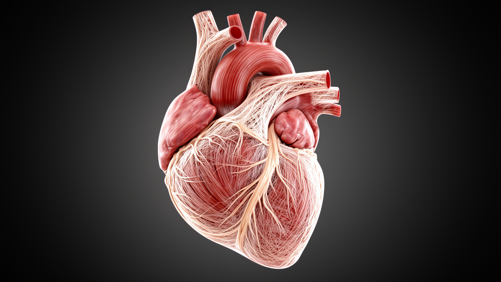
Published in European Radiology Experimental, the study provides a histology-validated framework for assessing these specialized cardiac conduction structures using relaxation-based MRI techniques. The work addresses a significant challenge in cardiac imaging: the inability of current in vivo MRI approaches to directly visualize thin, sparsely distributed Purkinje fibers. Instead, the study focuses on the collagen-rich extracellular matrix (ECM) that surrounds and supports these fibers, showing that specific MRI relaxation parameters are sensitive to this microstructural environment.
The cardiac conduction system (CCS) consists of specialized myocytes that initiate and propagate electrical impulses to coordinate ventricular activation. The terminal component of this system, the Purkinje fibers (PFs), forms a high-velocity conduction network essential for near-synchronous ventricular contraction. Structurally, these fibers are embedded within a highly organized extracellular matrix composed of subendocardial connective layers and false tendons (FTs). FTs are intracavitary fibromuscular bands predominantly composed of dense collagen bundles that surround and support PF bundles, providing structural stabilization and defining their anatomical trajectories. Because PFs are thin and sparsely distributed within the ventricular myocardium, they cannot be directly visualized using current in vivo MRI approaches. Instead, MRI relaxation contrast primarily reflects the composition and microstructural properties of the surrounding collagen-rich ECM associated with PF pathways.
The study aimed to evaluate whether qMRI could characterize these collagen-rich PF-associated pathways using histology as a structural ground truth. The research team utilized ex vivo porcine left ventricular specimens, selected because the pig heart’s ventricular geometry, myocardial fiber architecture, and Purkinje system organization closely resemble those of the human heart. Although pigs exhibit slightly more extensive subendocardial arborization, overall PF morphology and ECM-associated pathways are well conserved, supporting their use in ex vivo CCS studies. The specimens were obtained from five Finnish Landrace pigs, with tissue blocks dissected from the apical region of the left ventricular wall to preserve the anatomical orientation of PF-associated regions.
For the imaging protocol, the specimens were imaged at a clinically compatible 3-T field strength using an 18-channel knee coil. The team employed relaxation-based mapping techniques, including TRAFF (relaxation along a fictitious field), T1ρ (longitudinal rotating frame relaxation time), T2, and T1 mapping. These rotating-frame relaxation methods are sensitive to slow molecular motions and macromolecular environments, improving contrast in collagen-rich or structurally specialized myocardial regions. TRAFF is generated using amplitude- and frequency-modulated RAFF pulses, which improve clinical feasibility by reducing RF power deposition while demonstrating increased relaxation times in fibrotic myocardium. The in-plane resolution was 0.6 × 0.6 mm², with a slice thickness of 1.5 mm, and the total MRI acquisition time for each specimen was approximately 2–3 hours.
To establish a structural reference, the researchers processed corresponding Masson’s trichrome sections into optical density (OD) maps. This staining method enables clear visualization of collagen (blue) and myocardium (red), thereby outlining the fibrous scaffolds through which PFs course. Although PFs lack a specific histochemical marker, their distribution within collagen-rich substrates provides consistent anatomical landmarks for delineating their spatial organization. The histological image analysis involved decomposing RGB images into red and blue channels, converting them into OD maps, and segmenting tissue types based on characteristic staining patterns. Myocardium was identified from regions with predominant red channel intensity, collagen from regions with predominant blue channel intensity, and PF-associated regions from red-stained myocardial signal embedded within collagen-rich strands.
Histological analysis confirmed the presence of collagen-enriched PF-associated pathways within subendocardial tissue and false tendons. In the subendocardial region, PFs appeared as pale red myocytes embedded within collagen-rich strands beneath the endocardium. Within the FTs, PF bundles were similarly identified but enclosed by a denser collagen sheath and lined by an endothelial layer. Optical density-based segmentation reliably differentiated tissue components with low inter-section variability. The findings indicated that the differentiation of PF-associated regions was primarily driven by the surrounding collagen-rich ECM environment rather than intrinsic myocardial staining differences. PF-associated regions demonstrated OD_R values comparable to myocardium, while OD_B values were significantly lower than myocardium, highlighting the role of the collagen matrix in defining the structural signature.
Quantitative MRI results revealed consistent relaxation-time differences between high-collagen (HC) regions and remote myocardial control (MC) regions across the five cardiac samples. HC regions demonstrated markedly increased TRAFF values (214 ± 18 ms versus 119 ± 3 ms in MC, p < 0.001), T1ρ values (115 ± 24 ms versus 72 ± 3 ms, p < 0.05), and T2 relaxation times (71 ± 10 ms versus 52 ± 3 ms, p < 0.05) compared with MC. T1 values were also higher in HC than MC regions (1028 ± 36 ms versus 979 ± 43 ms, p < 0.05), although the relative contrast remained lower than that observed for rotating-frame relaxation parameters. These measurements suggest that relaxation-based MRI techniques are sensitive to the collagen-rich ECM substrates associated with Purkinje fibers.
The contrast between HC and MC regions was further quantified using the relative relaxation time difference (RRTD). RRTD analysis indicated that TRAFF and T1ρ provided the strongest relative contrast among the evaluated parameters. TRAFF demonstrated the highest RRTD (0.57 ± 0.08), followed by T1ρ (0.45 ± 0.23) and T2 (0.31 ± 0.15), whereas T1 exhibited the lowest relative contrast (0.05 ± 0.03). Pairwise comparisons showed that TRAFF RRTD was significantly greater than T2 and T1, with no significant difference between TRAFF and T1ρ. This hierarchy may reflect the differing sensitivities of these parameters to tissue microstructure, with rotating-frame methods being particularly sensitive to low-frequency molecular motions and macromolecular environments.
The study highlights that MRI sensitivity to PF-associated regions arises not from direct visualization of the fibers themselves, but from detecting their co-localized, collagen-rich ECM within FTs and subendocardial layers. This distinction is critical for interpreting relaxation-time contrast and underscores the role of ECM pathways as the primary MRI-visible substrates associated with PFs. The observed agreement between histological tissue characteristics and MRI relaxation behavior supports the interpretation that these relaxation-based imaging techniques are sensitive to PF-associated collagen-rich ECM substrates. This approach offers a non-invasive and potentially translatable means of probing PF-associated collagen substrates indirectly through their relaxation properties, leveraging sequences and spatial resolutions achievable on clinical 3-T scanners.
Despite these advances, several limitations should be considered. The sample size was modest, reflecting the exploratory feasibility nature of this ex vivo study, with ten sections analyzed from five hearts. Histological and OD analyses are inherently destructive and limited to ex vivo validation, and tissue deformation during histological processing may have influenced measurements in thin and heterogeneous PF-associated regions. Furthermore, although TRAFF and T1ρ showed increased sensitivity to collagen-rich PF-associated ECM pathways, these relaxation changes are not specific to PF-associated collagen and may also occur in other collagen-rich myocardial conditions, such as fibrosis or post-infarction scar tissue. All MRI data were acquired ex vivo, and no dedicated assessment of B0/B1-related artifacts was performed.
Translation of this approach toward in vivo imaging remains challenging and was beyond the scope of the present study. Future development will likely require improved spatial resolution, efficient acquisition strategies, and integration with anatomical landmarks or complementary structural MRI techniques to improve localization of PF-associated ECM pathways under clinical conditions. Current clinical MRI systems remain insufficient for direct visualization of individual PF structures, so future in vivo applications would likely rely on detecting PF-associated collagen-rich substrates rather than resolving microscopic PF architecture directly. Further optimization will be required to address cardiac motion, B0/B1 inhomogeneity, SAR constraints, and practical acquisition limitations.
Collectively, these findings support further investigation of non-invasive MRI approaches for characterizing conduction-related cardiac microstructure and ventricular conduction substrates. The study demonstrates the feasibility of MRI-based tissue characterization for studying conduction-related microstructure beyond the spatial resolution limits of conventional imaging. By establishing a histology-referenced framework, the research provides a foundation for future studies across species, disease models, and pathological substrates to evaluate the robustness and generalizability of the observed histology–MRI relationships. These efforts may support future investigation of conduction-related microstructure and ventricular conduction abnormalities using non-invasive MRI approaches.
Subject of Research: Quantitative MRI characterization of collagen-rich Purkinje fiber-associated pathways in ex vivo porcine hearts
Article Title: Histology-validated quantitative MRI characterization of collagen-rich Purkinje fiber-associated pathways in the ex vivo porcine heart
Article References: Li, Y., Casula, V., Pradeep, M., & Liimatainen, T. (2026). Histology-validated quantitative MRI characterization of collagen-rich Purkinje fiber-associated pathways in the ex vivo porcine heart. European Radiology Experimental, 10(1), Article 132. https://doi.org/10.1186/s41747-026-00793-0
Image Credits: AI Generated
DOI: 10.1186/s41747-026-00793-0
Keywords: Quantitative MRI, Purkinje fibers, Cardiac conduction system, Porcine heart, Ex vivo imaging, Collagen, TRAFF, T1ρ mapping, Histology, Cardiac microstructure, Histology-validated, quantitative
News Source: Ophelia Keating. (November 10, 2026). MRI Maps Collagen Pathways in Porcine Hearts. Scienmag.