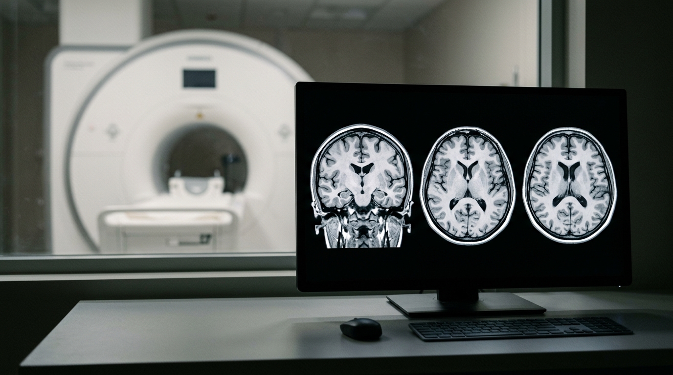
A machine-learning model analyzing structural magnetic resonance imaging scans classified older adults with mild cognitive impairment from cognitively normal individuals with high internal accuracy, according to a study using Alzheimer’s Disease Neuroimaging Initiative data. The retrospective cross-sectional evaluation examined neuroimaging data alongside basic demographic characteristics. By combining discrimination metrics with calibration assessments and post hoc feature interpretations, the research team developed a comprehensive framework for automated cognitive classification.
Prior research utilizing machine learning on structural magnetic resonance imaging has repeatedly shown potential for distinguishing mild cognitive impairment from normal cognition. However, earlier studies frequently emphasized discrimination alone, leaving model calibration, comparative evaluation across different algorithms, and anatomically interpretable contributions less consistently integrated. Furthermore, researchers noted that identifying influential structural imaging features in an algorithmic pipeline does not establish their independent biological significance or confirm them as disease mechanisms.
To address these gaps, researchers analyzed records from 2,649 participants aged over 65 years from the Alzheimer’s Disease Neuroimaging Initiative. The cohort comprised 1,285 cognitively normal individuals and 1,364 participants with mild cognitive impairment. Using least absolute shrinkage and selection operator regression in the training set, the team selected eighteen structural morphometric features, which were then combined with three demographic variables: age, sex, and educational attainment.
The researchers compared five distinct algorithms using an internally held-out test set: logistic regression, support vector machine, random forest, Light Gradient Boosting Machine, and extreme gradient boosting, known as XGBoost. Model performance across all systems was assessed using discrimination, classification metrics, calibration, and decision-curve analysis. In addition, Shapley Additive Explanations were implemented to provide post hoc model interpretation of anatomically relevant feature contributions.
Among the evaluated algorithms, the XGBoost-based final model achieved the strongest classification performance on the internally held-out test set. The final model reached an area under the receiver operating characteristic curve of 0.917, with a 95 percent confidence interval of 0.889 to 0.945. By comparison, logistic regression achieved an area under the curve of 0.865, with a 95 percent confidence interval of 0.836 to 0.894, demonstrating a statistically significant difference under paired DeLong testing with a P value of 0.048.
Detailed classification metrics further characterized the diagnostic performance of the final gradient boosting system on the test data. The XGBoost model demonstrated an accuracy of 0.847, a sensitivity of 0.854, and a specificity of 0.839.
Post hoc interpretation using Shapley Additive Explanations identified the most influential anatomical features guiding the classification. Bilateral inferior lateral ventricular volumes, bilateral hippocampal volumes, and left middle temporal cortical thickness contributed most heavily to the model’s predictions. The investigators emphasized that these importance rankings describe mathematical contributions to algorithmic predictions rather than independently established biomarkers or verified underlying disease mechanisms.
The investigators concluded that a structural magnetic resonance imaging framework incorporating demographic data demonstrated good internal discrimination between cognitively normal and mildly impaired participants in the study cohort. However, the authors noted these findings represent cohort-specific classification evidence, emphasizing that independent external validation is required before any potential clinical application.
Subject of Research: Machine learning classification of mild cognitive impairment using structural MRI
Article Title: Structural MRI-based machine learning classification of mild cognitive impairment in the Alzheimer’s Disease Neuroimaging Initiative
Article References: Wu, L., Dong, Y.-T., Sun, X.-X., Chen, Y., Luo, X., Mu, X., & Chen, Z.-J. (2026). Structural MRI-based machine learning classification of mild cognitive impairment in the Alzheimer’s Disease Neuroimaging Initiative. BMC Geriatrics. https://doi.org/10.1186/s12877-026-08701-2
Image Credits: AI Generated
DOI: 10.1186/s12877-026-08701-2
Keywords: structural MRI, machine learning, mild cognitive impairment, Alzheimer's Disease Neuroimaging Initiative, XGBoost, logistic regression, support vector machine, random forest, LightGBM, LASSO regression, hippocampal volume, inferior lateral ventricles, middle temporal cortex, Shapley Additive Explanations, Brier score, decision-curve analysis, model calibration
News Source: Teresa Odom. (November 18, 2026). MRI Machine Learning Model Accurately Identifies Mild Cognitive Impairment in Cohort. Scienmag.